❌

Reading view

Ultrasound waves that jiggle deep brain structures may relieve delusions in schizophrenia, small trial hints

Stimulating the brain with ultrasound could help counter the delusions people experience in schizophrenia, a small trial finds.

The trial involved transcranial ultrasound stimulation, in which a device placed on a patient's head delivers ultrasound waves through the skull and into the brain. Its goal is to change the activity in a targeted brain region.

Delusions are false beliefs that are very difficult to shake; for example, a person might believe they are being stalked when they aren't or think that a major disaster is about to occur without any evidence to suggest so. In the trial, researchers found that a 10-minute session of the ultrasound treatment reduced the severity of delusions for at least a week in patients with schizophrenia or a related disorder.

This targeted ultrasound treatment delivered directly to the brain could avoid some of the full-body effects of antipsychotic medications, such as weight gain and high blood pressure. That said, the new results, published Tuesday (Oct. 20) in the journal npj Acoustics, are a first step and will need further verification in larger trials.

"It's too early to know for certain what the therapeutic benefits will be, but it's very exciting," study co-author Dr. Donald Goff, a professor of psychiatry at the New York University Grossman School of Medicine, told Live Science.

Targeting deep brain structures

The pilot study included 10 patients with either schizophrenia or schizoaffective disorder. In both conditions, patients experience psychotic symptoms, including delusions and hallucinations. Schizoaffective disorder also includes mood symptoms, such as mania.

The researchers interviewed each participant to characterize the content of their delusions, and also documented their cognitive state and experiences with auditory hallucinations β€” perceptions of sounds that aren't actually occurring. The participants then received both an ultrasound treatment and a placebo treatment, spaced one week apart.

Other noninvasive therapies β€” such as transcranial magnetic stimulation, which uses magnetic fields β€” can also stimulate the brain. But the ultrasound technique differs from these therapies because ultrasound waves can penetrate deeper into the brain with greater specificity, study co-author Shy Shoham, a neural engineer at NYU Grossman, told Live Science. He compared the way a transducer directs ultrasound waves toward a given brain region with the way a lens focuses light.

In this case, the researchers focused this lens on a brain network called the cortico-striatal-pallido-thalamic circuit, which includes structures underneath the brain's outer cortex. The relationship between this circuit and schizophrenia symptoms is still somewhat unclear, partly because its deeper components are hard to study in adequate resolution with MRI, said co-author Dr. Wei Qi, a clinical assistant professor of psychiatry at NYU Grossman. However, some past studies have suggested that this network plays a part in schizophrenia.
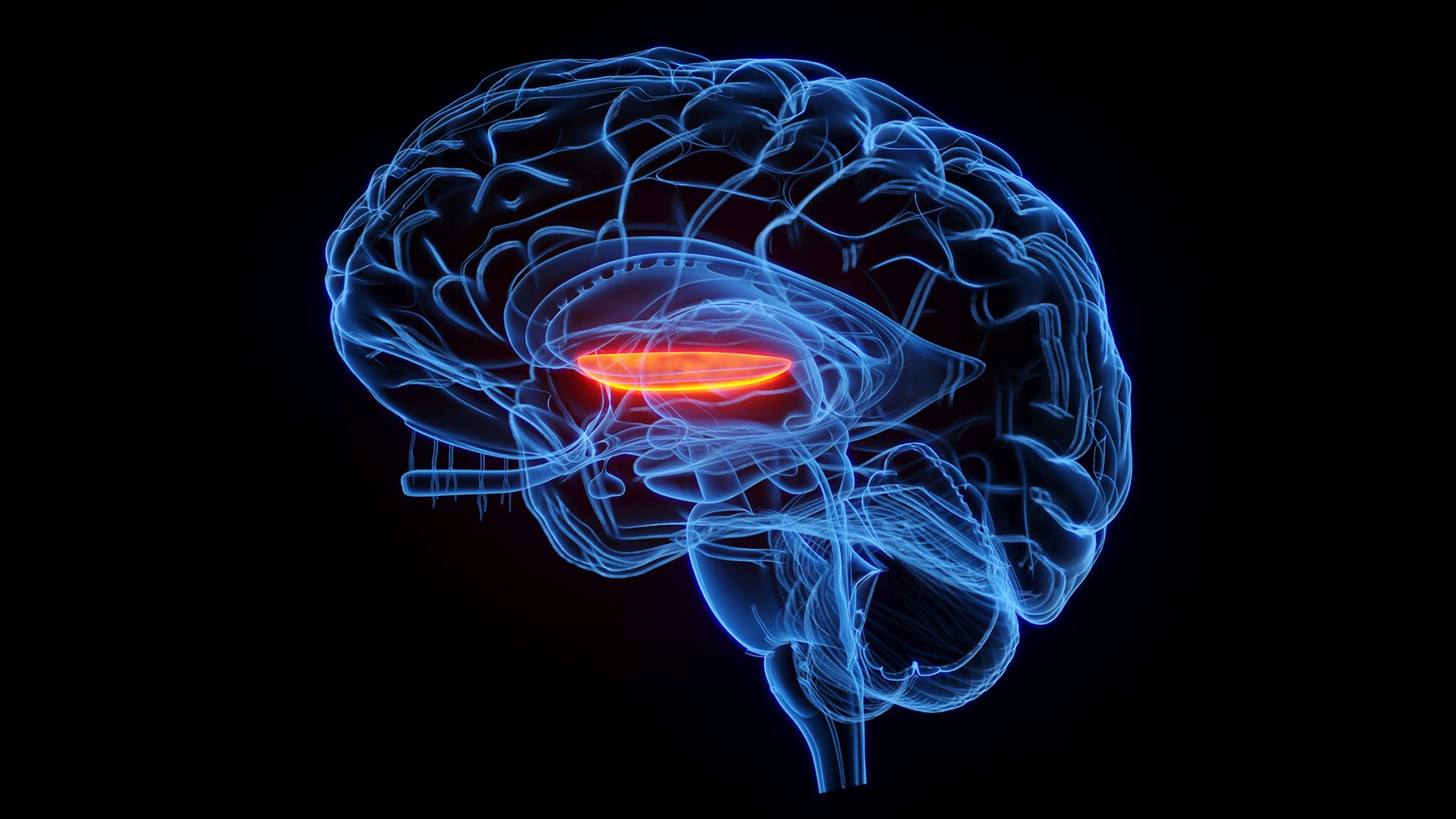
A cartoon of a transparent brain with an inner region glowing red.

The ultrasound treatment targeted a brain structure called the globus pallidus internus (orange). (Image credit: SEBASTIAN KAULITZKI/SCIENCE PHOTO LIBRARY via Getty Images)

Much of what scientists know about the network comes from research in movement disorders, such as Parkinson's disease, in which there is an imbalance between excitation and inhibition in the neurons that control movement. A similar dynamic could help to explain symptoms of schizophrenia, the scientists think.

"Our hypothesis is that, in schizophrenia, there is an imbalance between excitation and inhibition regarding what in the environment is actually important," Qi told Live Science. "The failure to inhibit irrelevant signals could be the basis of psychosis."

"Quite optimistic"

The researchers surveyed the study participants after each treatment, asking about the degree of belief each person had in their delusions and the amount of stress they caused. Compared with the placebo treatment, the ultrasound stimulation was associated with less-severe delusions over the following week. There was no significant change in patients' auditory hallucinations or overall cognition, though.

The ultrasound therapy came with mild side effects, such as fatigue and headache. Some patients had transient increases in delusions, which Qi attributed to normal fluctuations in symptoms that are expected in schizophrenia. These outcomes were just as common after the placebo treatment, so they may not be related to the ultrasound specifically.

The stimulation was aimed specifically at the right side of a brain structure called the globus pallidus internus (GPI). In a previous study, the authors found that greater connectivity between this region and another structure β€” the dorsal anterior cingulate gyrus β€” was linked to more-severe psychotic symptoms in schizophrenia. So, they made the GPI their target.

To date, "there haven't been many successful therapeutic attempts at targeting some of these deeper structures," said Dr. Deepak Sarpal, an associate professor of psychiatry at the University of Pittsburgh School of Medicine who was not involved with the study. Ultrasound waves reach brain regions under the cortex in a less-invasive manner than deep brain stimulation, in which a surgeon implants an electrode in a patient's brain, Sarpal noted.

The researchers are not the only group using transcranial ultrasound to influence the cortico-striatal-pallido-thalamic circuit. Earlier this year, another group stimulated portions of the striatum, which is located directly under the cortex, in patients with schizophrenia. They evaluated hallucinations rather than delusions, finding that the treatment reduced symptoms for at least a week after the procedure.

In this line of research, the targeted nature of transcranial ultrasound presents advantages as a research tool as well as a treatment approach.

"If we find that a specific circuit or a node in a circuit seems to be closely linked to symptoms we're trying to treat, then it will be a matter of understanding what pattern of stimulation may allow us to correct that," Goff said. Qi, Goff and colleagues now plan to continue testing their technique on more patients and to experiment with different doses of ultrasound.

"A lot more work needs to be done," Goff said, "but we're really quite optimistic about its promise."

This article is for informational purposes only and is not meant to offer medical advice.

See how much you know about the most complex organ in the human body with our brain quiz!

  •  

Teenager's rare autoimmune disorder caused her to hallucinate a flying green dinosaur

The patient: A 16-year-old girl in California

The symptoms: The teenager was referred to an emergency department because she was having hallucinations, including seeing a flying green dinosaur and hearing voices telling her to hurt others. She also had a sense that she was being followed.

In the emergency room, she was trembling and sweating and had a racing pulse and shallow, fast breathing. She did not have a fever, although she did have one at the hospital that had referred her.

During her conversations with ER clinicians, the teen reacted to the vision of the dinosaur as though the creature were present in the room. She reported that she had experienced these hallucinations since seventh grade,‬ so for about four years.

While her parents countered that she hadn’t exhibited psychotic symptoms until 48 hours before they arrived in the ER, they noted that her interest in school and extracurriculars had waned over the past month.

What happened next: Because of the patient's abnormal vital signs, the ER doctors suspected that her hallucinations might be secondary to some physical ailment, such as trauma to the brain, they wrote in a case report. However, a CT scan showed no signs of swelling or hemorrhage in her head. The doctors attempted a lumbar puncture (also called a spinal tap) but failed to collect any cerebrospinal fluid that they could test for irregularities.

The doctors judged the girl to be too medically unstable to be referred to the hospital's psychiatric ward, so she was admitted to the general pediatric department. Pediatric neurologists suggested an electroencephalogram (EEG), a test that measures electric signaling in the brain, but this revealed no further clues.

On her second day in the hospital, the girl entered a complete catatonic state. Her specific symptoms were not described in the report, but in general, catatonia disrupts how a person processes and reacts to the world around them. Catatonic patients often stop moving and speaking. Doctors treated the patient with lorazepam, which slows down nervous-system signaling, and her catatonia subsided in days.

Meanwhile, interventional radiologists performed a successful second lumbar puncture, but the girl's cerebrospinal fluid appeared normal in the initial panels they ran.

After the patient had spent eight days in the hospital, psychiatrists recommended that the girl be transferred to their ward. However, this was not possible because she developed problems with feeding herself. Her continued psychotic symptoms were treated with the antipsychotic drug olanzapine.

The diagnosis: While initial tests of the girl's cerebrospinal fluid did not show signs of meningitis or other abnormalities, a later test came back positive for autoantibodies β€” immune proteins that target healthy tissues instead of foreign invaders. These antibodies targeted neuronal intermediate filament (NIF), a protein that helps give nerve cells structural support.

Based on this finding, the patient was diagnosed with autoimmune encephalitis, a condition in which the body produces antibodies that attack nervous tissue, leading to harmful inflammation that can cause psychiatric symptoms.

Autoimmune encephalitis is often triggered by a tumor; proteins produced by the growth can resemble proteins within nervous tissue, and the immune system develops a harmful response against the latter while attempting to fight the tumor. The doctors performed MRIs and a PET scan to look for tumors, but they did not find any.

Illustration of Y shaped antibody proteins

The girl was diagnosed with autoimmune encephalitis, a condition in which antibodies attacked her own nervous tissue. (Image credit: Shutterstock)

The treatment: The girl received intravenous infusions of a steroid to reduce inflammation, as well as immunoglobulin β€” antibodies from healthy donors that are often used to calm autoimmune reactions. She also got an infusion of rituximab, a drug that kills B lymphocytes, a type of white blood cell that makes antibodies like the ones attacking the girl's neurons.

In addition to these infusions, the girl underwent five sessions of plasmapheresis over the next month. In that procedure, blood is drawn from the body, a machine separates plasma (the liquid portion of blood) from blood cells, and the extracted plasma is substituted with a replacement fluid before that blood is returned to the patient. This treatment can help rid the body of autoantibodies.

The girl was discharged after having spent almost two months in the hospital. She was treated with ongoing prescriptions of olanzapine and lorazepam, as well as prednisone and monthly immunoglobulin treatments. As her symptoms improved over the following year, she stopped taking the psychiatric drugs but continued her immunotherapy.

What makes the case unique: Autoimmune encephalitis is rare. One study conducted in the U.K. suggests it affects only 5 to 8 out of every 100,000 people. Additionally, most data about the condition in pediatric patients comes from cases caused by autoantibodies that target NMDA receptors, which detect a key chemical messenger in the brain, rather than NIF.

The case report did not name a specific trigger for the girl's condition, but the authors noted that anti-NIF autoimmune encephalitis has been documented in adults following infections, including certain tick-borne bacterial infections, HIV and COVID-19. Cases associated with these infections are rarer than those linked to tumors.

The doctors did not attribute the teen's case to a specific infection, but her family did report that she'd had a cough three weeks before going to the emergency room. The family had visited the Philippines, and the girl and multiple relatives came down with the same illness.

For more intriguing medical cases, check out our Diagnostic Dilemma archives.

This article is for informational purposes only and is not meant to offer medical advice.

Can you guess the diagnosis in these strange medical cases? Find out with our diagnostic dilemma quiz!

  •  

Jonathan the tortoise has lived to 194 years old β€” now scientists think they know why

The remote south Atlantic island of St. Helena boasts an international celebrity: the tortoise Jonathan, the oldest living land animal. At 194 years old, he has outlasted the expected lifespan for an Aldabra giant tortoise (Aldabrachelys gigantea) by about a century. Now, scientists have identified a range of unique genetic and epigenetic traits that may hold the key to this longevity.

The new research zoomed in on Jonathan's genes, as well as the chemical tags that sit on top of DNA molecules, known as "epigenetic markers." These markers help control which genes are activated and to what degree. The analysis finds that Jonathan's genes associated with mitochondrial function are in great shape, epigenetically speaking, which suggests that they've played a big role in the tortoise's success.

While mitochondrial function has long been linked to longevity and mitochondrial dysfunction has been tied to disease, Jonathan offers fresh insight into that dynamic, said study co-author Stephen Clark, chief scientist of the Kallel Foundation, a nonprofit in Nashville researching drug targets to promote human longevity.

Clark and colleagues published their findings Wednesday (Oct. 7) in the journal Science Advances.

Decoding Jonathan's secrets to long life

Nearly a decade ago, the researchers enlisted Joe Hollins, the vet tasked with Jonathan's care, to collect samples from the record-setting animal. Island authorities forbade the vet from drawing the tortoise's blood, fearing the risk of an infection.

"I didn't want to be the doctor that killed Jonathan," Clark told Live Science. Hollins collected samples via cheek swab instead.

When the samples arrived in the U.S., the researchers sequenced them, but when their computers kept crashing, they discovered that the DNA had been isolated from bacteria in Jonathan's mouth rather than the tortoise's own cells.

Clark "had to go back and beg" for permission to collect more data, he said. The vet then sent cheek scrape samples, which are collected with a slightly different tool than cheek swabs. This time, the DNA was in fact from Jonathan.

The DNA isolated from those cheek tissue samples was more fragmented than it would have been in a blood sample, so the researchers filled in the gaps using an existing reference genome from a 36-year-old Aldabra tortoise named Tank. They also compared Jonathan's DNA to that of a GalΓ‘pagos tortoise (Chelonoidis abingdoni) named Lonesome George, who was believed to be over 100 years old at the time of his death in 2012.

The team found that, compared to Tank and Lonesome George, Jonathan carried 287 unique variants of genes that had previously been linked to aging-related pathways. These included genes involved in DNA repair and the function of telomeres β€” the protective tips at the end of chromosomes that tend to shorten with age.

These pathways are also related to aging processes in humans, highlighting a "broader signature of aging" across species, Greer Dolby, an assistant professor of biology at The University of Alabama at Birmingham, who was not involved with the study, told Live Science.

The researchers also looked at epigenetic changes, including DNA methylation, which is when a chemical tag called a methyl group binds the DNA. Certain patterns of DNA methylation can function as a "clock" in that they reflect an organism's biological age; scientists have recently harnessed these patterns in the study of human aging. In this case, the team compared the DNA methylation across Jonathan's genome to that of young and old Aldabra tortoises.

A large tortoise sits on a grassy lawn.

Researchers compared DNA from Jonathan (pictured) to that of other Aldabra giant tortoises. (Image credit: Kevin Gepford, CC BY-SA 4.0)

The methylation patterns were more disordered in the older tortoises, reflecting random changes acquired over time, the researchers found. Scientists call this phenomenon "methylation entropy." For most genes, Jonathan's DNA methylation patterns were more disordered than those of the young tortoises. But in a suite of genes tied to mitochondrial function β€” specifically the bits of those genes that switch them "on" β€” Jonathan had more organized methylation patterns. That might keep expression of these genes consistent over time, the researchers theorized.

Mitochondria produce energy that cells need to function properly, repair themselves and reduce further damage to DNA. "Keeping the entropy low in these mitochondrial genes, or keeping pristine mitochondria, is likely to be a contributor to longevity," Clark said.

However, the authors couldn't determine a causal relationship β€” they didn't run any tests that could definitively prove that Jonathon's lack of entropy in these genes explains his long life. Clark argued that additional experiments on a blood sample from Jonathan could offer a more comprehensive view of his genome.

Jonathan's full genetic profile can't be known until after the tortoise's death, when researchers can collect DNA from more tissues, said Vincent Lynch, a professor of biology at the University at Buffalo, who was not involved with the study.

"Some organs are more susceptible to different diseases of old age than other ones, and that happens because mutations accumulate over time," he told Live Science. "We would ideally like to know what those mutations are in those specific tissues."

Furthermore, Lynch questioned whether entropy was a useful characterization of age-related DNA methylation patterns, as methylation can be unpredictable. That said, he agreed that the genes flagged in the study offer helpful data for future work on longevity.

More experiments are needed to determine whether these mitochondrial genes actually play a role in longevity across all Aldabra tortoises, let alone in other animals, or if Jonathan is just an outlier.

"Maybe Jonathan is just really good at being old," Lynch said.

  •  

Woman 'saw dead people' and fired a gun at them after taking too many aspirin

The patient: A 65-year-old woman in Michigan

The symptoms: The woman arrived at the emergency room via ambulance and asked to see a psychiatrist for her visual hallucinations. "I'm seeing dead people," she told clinicians, adding that she'd even fired a gun at her hallucinations in one instance, according to a report of her case. (The report does not describe what the hallucinations looked like.)

Despite this reaction to her hallucinations, she was calm and alert during her physical exam, with normal vital signs other than high blood pressure. She reported a history of high blood pressure, diabetes, heart failure and chronic back pain but no prior psychiatric history.

What happened next: Since the woman had no past psychiatric history, doctors opted for a medical assessment before consulting a psychiatrist. A blood test revealed mild metabolic acidosis, meaning her blood was too acidic. A toxicology screen detected high levels of salicylates, a class of compound that includes acetylsalicylic acid, better known as aspirin.

The diagnosis: When questioned, the woman acknowledged that she had been taking over 15 aspirin pills a day for multiple weeks. Each of these pills, marketed for back and body pain, contained 500 milligrams of aspirin.

Upon further questioning, the woman confirmed that she had tinnitus (ringing in her ears), which is a classic symptom of salicylate toxicity. In other words, she'd overdosed on aspirin.

The treatment: Doctors consulted experts at Michigan's poison control center, who recommended intravenous (IV) treatment with sodium bicarbonate (the chemical name for baking soda). This treatment alkalinizes blood and urine, meaning it makes them less acidic. This enables more salicylate to be excreted through the kidneys, allowing the drug to be eliminated through the urine.

After 11 hours of the IV treatment, the woman's blood salicylate levels dropped from 28.8 milligrams per deciliter (mg/dL) to 18.3 mg/dL. Her hallucinations ceased and the tinnitus improved within 48 hours of her arrival at the hospital. She was discharged without having seen a psychiatrist.

Close-up photo of a woman's ear

Ringing in the ear is a common symptom associated with aspirin overdose. (Image credit: liza5450/Getty Images)

What makes the case unique: Aspirin is generally safe when used in doses indicated on the bottle or advised by a person's doctors. However, taking too much aspirin can irritate the gastrointestinal tract, as well as disrupt the activity of parts of the nervous system.

At high enough doses, salicylates can stimulate part of the brain called the medulla, which regulates breathing. This results in hyperventilation that causes the body to lose too much carbon dioxide, and at first, the blood becomes more alkaline. This process is known as respiratory alkalinosis.

However, salicylates also disrupt metabolism, leading to metabolic acidosis from a buildup of lactic acid in the body, as the woman experienced. When metabolic acidosis overwhelms the respiratory alkalinosis and causes the blood to acidify, organ damage can occur. These metabolic effects may also explain the tinnitus, as it drives increases in arachidonic acid that stimulate receptors in the inner ear.

The woman's daily intake of roughly 75 milligrams of aspirin per kilogram of body weight was about half the typical dose associated with acute salicylate poisoning. However, it was still a large amount to take every day for weeks, and older age is also a risk factor for severe outcomes of acute salicylate toxicity.

The authors of the case report noted that although she had tinnitus, she lacked many other hallmark symptoms of salicylate overdose, such as nausea and fever.

To the clinicians, the woman's case served as a reminder that long-term aspirin toxicity may result in different symptoms than acute poisoning does. In past case reports, other patients with chronic salicylate toxicity have also reported visual hallucinations. But that potential symptom is not as well understood as more common symptoms like tinnitus.

The case report authors added that longer-term exposure to aspirin may be associated with more neurological symptoms because chronic exposure increases the odds of the drug crossing the blood-brain barrier β€” a gatekeeper that allows only certain substances from the blood into the brain. They added that doctors should consider poisoning, including salicylate toxicity, for patients with psychotic symptoms with no clear cause.

For more intriguing medical cases, check out our Diagnostic Dilemma archives.

This article is for informational purposes only and is not meant to offer medical advice.

Can you guess the diagnosis in these strange medical cases? Find out with our diagnostic dilemma quiz!

  •  

Diagnostic dilemma: A woman got a lung transplant β€” and ended up with a peanut allergy, too

The patient: A 47-year-old woman in Canada

The symptoms: The woman underwent a lung transplant to treat interstitial lung disease, which involves harmful inflammation and scarring of lung tissue. While recovering in the hospital, she ate a bowl of breakfast cereal, which activated a nearly two-hour episode of flushing, itching, shortness of breath and low blood pressure.

This severe, life-threatening allergic reaction is known as anaphylaxis. A report of the woman's case does not mention specific treatment she received for this reaction, but it notes that it resolved after two hours.

(Treatment for anaphylaxis involves epinephrine (also called adrenaline), and it can also require antihistamines, cortisone, oxygen and beta-agonists, such as albuterol, which help improve breathing.)

What happened next: The woman experienced the same symptoms in the weeks after being discharged from the hospital β€” after eating a coconut cream pie and after having an almond candy bar.

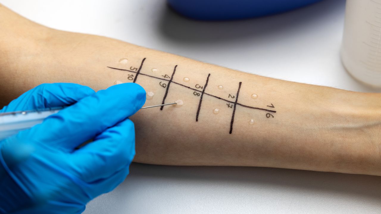
The diagnosis: Two months after her lung transplant, the woman underwent a skin-prick allergy test, which revealed a sensitivity to peanut and pine nut allergens. She did not have an allergic response to tree nuts, such as the almonds in the candy bar, nor to coconut. None of the foods that triggered her symptoms were known to contain peanuts, but none were certified as peanut-free, either.

Notably, the woman's lung donor had been a 12-year-old boy with a known peanut allergy but no history of anaphylaxis. Her doctors theorized that the transplanted lung tissue contained immune cells from the donor that were sensitized to nut allergens.

The treatment: The woman did not receive a specific treatment to get rid of this new allergic response. However, the sensitivity subsided in follow-up skin-prick tests, and they had disappeared completely by a year after her surgery. She ate peanuts in a supervised setting a few months after the one-year mark and had no reaction.

The doctors reasoned that the sensitized immune cells from the donor must have cleared from her body.

What makes the case unique: Only one other case of peanut allergy following a lung transplant had been documented before the woman's case was published in 2011.

Scientists had only recognized the risk of allergies being passed from organ donors to recipients a little over a decade before the report, according to a 2025 review of such instances. Doctors had previously reported allergy transfer linked to bone marrow transplants, which makes sense given that bone marrow produces immune cells. But the first known donor-acquired allergy from a solid-organ transplant was observed in 1997 following a combined kidney-liver transplant from a donor who was allergic to peanuts.

The authors of the 2025 review noted that donor-acquired allergies are more commonly associated with lung and liver transplants than with transplants of other solid organs. That could be because these organs are rich in a type of immune cell that produces allergen-specific antibodies, called B lymphocytes.

Other potential culprits for donor-acquired allergies are mast cells and basophils, two types of immune cell that bind to those allergen-specific antibodies. The cells hold onto the antibodies, and when they encounter an allergen, they release the inflammatory compounds responsible for anaphylaxis.

In the woman's case, a blood test for peanut-specific antibodies came back negative. This suggested that her body was not continually generating the antibodies. That could serve as possible evidence that her reaction was caused by antibodies bound to mast cells or basophils within the donor tissue, rather than antibodies produced by lymphocytes.

That said, the woman's reaction to the skin-prick test could imply that lymphocytes were still involved in the allergic reaction, the doctors added, as they did not know whether mast cells or basophils could migrate to the skin the way circulating antibodies could.

Regardless of the cell type behind the woman's peanut allergy, the sensitivity faded after a year, consistent with other cases described in the 2025 review. Still, the review authors called for better donor allergy labels to inform allergy testing and treatment for organ recipients. The doctors in the case report came to a similar conclusion, noting that detailed donor allergy histories and skin-prick testing after organ transplant could help identify donor-acquired allergies.

For more intriguing medical cases, check out our Diagnostic Dilemma archives.

This article is for informational purposes only and is not meant to offer medical advice.

  •  
❌