Login
Scientists Used a Magic Mushroom Compound to Calm an Extremely Aggressive Fish
20 October 2026 at 20:27
 Scientists discovered that psilocybin can make one of nature's most aggressive fish surprisingly mellow.
 Scientists discovered that psilocybin can make one of nature's most aggressive fish surprisingly mellow.

Stimulating the brain with ultrasound could help counter the delusions people experience in schizophrenia, a small trial finds.
The trial involved transcranial ultrasound stimulation, in which a device placed on a patient's head delivers ultrasound waves through the skull and into the brain. Its goal is to change the activity in a targeted brain region.
Delusions are false beliefs that are very difficult to shake; for example, a person might believe they are being stalked when they aren't or think that a major disaster is about to occur without any evidence to suggest so. In the trial, researchers found that a 10-minute session of the ultrasound treatment reduced the severity of delusions for at least a week in patients with schizophrenia or a related disorder.
This targeted ultrasound treatment delivered directly to the brain could avoid some of the full-body effects of antipsychotic medications, such as weight gain and high blood pressure. That said, the new results, published Tuesday (Oct. 20) in the journal npj Acoustics, are a first step and will need further verification in larger trials.
"It's too early to know for certain what the therapeutic benefits will be, but it's very exciting," study co-author Dr. Donald Goff, a professor of psychiatry at the New York University Grossman School of Medicine, told Live Science.
The pilot study included 10 patients with either schizophrenia or schizoaffective disorder. In both conditions, patients experience psychotic symptoms, including delusions and hallucinations. Schizoaffective disorder also includes mood symptoms, such as mania.
The researchers interviewed each participant to characterize the content of their delusions, and also documented their cognitive state and experiences with auditory hallucinations — perceptions of sounds that aren't actually occurring. The participants then received both an ultrasound treatment and a placebo treatment, spaced one week apart.
Other noninvasive therapies — such as transcranial magnetic stimulation, which uses magnetic fields — can also stimulate the brain. But the ultrasound technique differs from these therapies because ultrasound waves can penetrate deeper into the brain with greater specificity, study co-author Shy Shoham, a neural engineer at NYU Grossman, told Live Science. He compared the way a transducer directs ultrasound waves toward a given brain region with the way a lens focuses light.
In this case, the researchers focused this lens on a brain network called the cortico-striatal-pallido-thalamic circuit, which includes structures underneath the brain's outer cortex. The relationship between this circuit and schizophrenia symptoms is still somewhat unclear, partly because its deeper components are hard to study in adequate resolution with MRI, said co-author Dr. Wei Qi, a clinical assistant professor of psychiatry at NYU Grossman. However, some past studies have suggested that this network plays a part in schizophrenia.
Much of what scientists know about the network comes from research in movement disorders, such as Parkinson's disease, in which there is an imbalance between excitation and inhibition in the neurons that control movement. A similar dynamic could help to explain symptoms of schizophrenia, the scientists think.
"Our hypothesis is that, in schizophrenia, there is an imbalance between excitation and inhibition regarding what in the environment is actually important," Qi told Live Science. "The failure to inhibit irrelevant signals could be the basis of psychosis."
The researchers surveyed the study participants after each treatment, asking about the degree of belief each person had in their delusions and the amount of stress they caused. Compared with the placebo treatment, the ultrasound stimulation was associated with less-severe delusions over the following week. There was no significant change in patients' auditory hallucinations or overall cognition, though.
The ultrasound therapy came with mild side effects, such as fatigue and headache. Some patients had transient increases in delusions, which Qi attributed to normal fluctuations in symptoms that are expected in schizophrenia. These outcomes were just as common after the placebo treatment, so they may not be related to the ultrasound specifically.
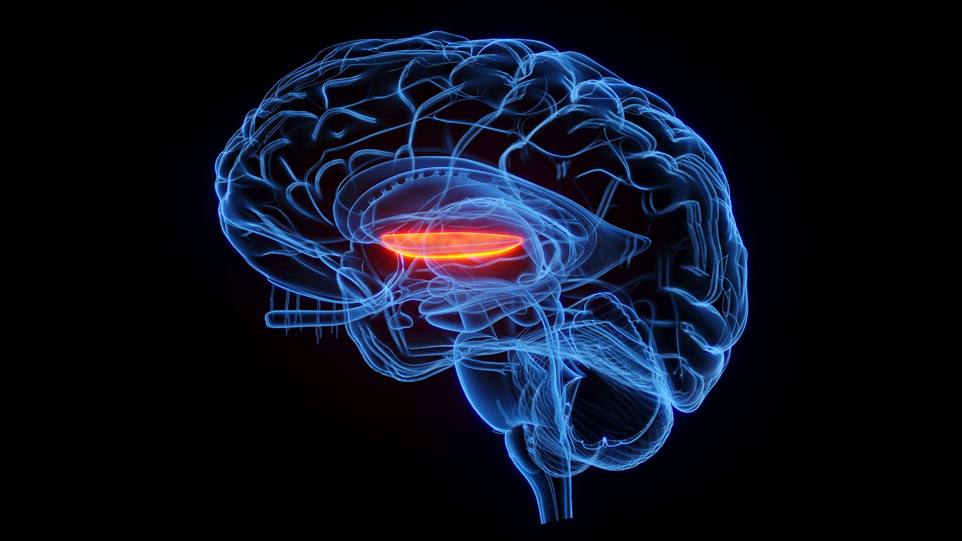
The stimulation was aimed specifically at the right side of a brain structure called the globus pallidus internus (GPI). In a previous study, the authors found that greater connectivity between this region and another structure — the dorsal anterior cingulate gyrus — was linked to more-severe psychotic symptoms in schizophrenia. So, they made the GPI their target.
To date, "there haven't been many successful therapeutic attempts at targeting some of these deeper structures," said Dr. Deepak Sarpal, an associate professor of psychiatry at the University of Pittsburgh School of Medicine who was not involved with the study. Ultrasound waves reach brain regions under the cortex in a less-invasive manner than deep brain stimulation, in which a surgeon implants an electrode in a patient's brain, Sarpal noted.
The researchers are not the only group using transcranial ultrasound to influence the cortico-striatal-pallido-thalamic circuit. Earlier this year, another group stimulated portions of the striatum, which is located directly under the cortex, in patients with schizophrenia. They evaluated hallucinations rather than delusions, finding that the treatment reduced symptoms for at least a week after the procedure.
In this line of research, the targeted nature of transcranial ultrasound presents advantages as a research tool as well as a treatment approach.
"If we find that a specific circuit or a node in a circuit seems to be closely linked to symptoms we're trying to treat, then it will be a matter of understanding what pattern of stimulation may allow us to correct that," Goff said. Qi, Goff and colleagues now plan to continue testing their technique on more patients and to experiment with different doses of ultrasound.
"A lot more work needs to be done," Goff said, "but we're really quite optimistic about its promise."
This article is for informational purposes only and is not meant to offer medical advice.
See how much you know about the most complex organ in the human body with our brain quiz!
Recent research challenges a long-standing idea about how the brain develops: Rather than arising from a single source, the brain may emerge from two distinct populations of cells, the study finds.
"The brain appears as one organ," said study co-author Kyle Loh, an associate professor of developmental biology at Stanford University. "However, we discovered that the front part of the brain and the back part of the brain come from different sources, which are pushed together to create the appearance of a single organ," Loh told Live Science in an email.
The findings, published in September in the journal Nature Neuroscience, not only challenge established models of how the brain forms but may also explain why researchers have struggled to grow certain types of brain cells in the laboratory. The scientists say the work could offer new ways to investigate diseases that affect the brainstem, the "stalk" that connects the brain to the spinal cord.
The human brain is divided into three main regions — called the forebrain, midbrain and hindbrain — that perform different but interconnected functions.
The forebrain, which includes the cerebral hemispheres, is involved in higher-level cognitive processes, such as language, planning and abstract thinking. The midbrain, the uppermost part of the brainstem, connects the forebrain and the hindbrain and acts as a key relay center for sensory information and movement control. The hindbrain — which includes the bottom of the brainstem, along with other structures — also helps to coordinate movement, as well as regulate essential functions such as breathing, sleep and heart rate.
Historically, the hindbrain has been difficult to study. One reason is that scientists have faced persistent challenges in generating functional hindbrain neurons in laboratory conditions.
To help understand why, Loh and his team looked at the early stages of embryonic development in lab mice. They focused on gastrulation, the point when cells begin to organize into structures that will later form different organs. The researchers identified two distinct populations of immature cells that would go on to give rise to nervous system tissue, including the brain.
One cell population expressed a gene called Otx2, which helps to direct the early development of the forebrain and midbrain. The second group of cells expressed the gene Gbx2, which is involved in the development of the midbrain and hindbrain.
The functions of these genes were already known, but their distinct expression in these two groups of cells was revealed by the new research. The cell populations remained distinct as they developed into full-fledged nervous tissue, rather than overlapping.
The team also examined chromatin, the molecular structure that packages DNA tightly inside the nucleus and helps regulate which genes a cell can use. The two cell populations show different chromatin configurations, the team found, which further supports the idea that they follow separate developmental pathways.
That may help to explain why scientists' attempts to grow hindbrain neurons have been largely unsuccessful.
"Previous attempts to make hindbrain neurons likely tried to coax forebrain and midbrain progenitors into hindbrain cells, which our study shows is not possible," study co-author Rayyan Jokhai, a graduate student in Loh's lab, said in a statement.
Using the information uncovered in their experiments, the researchers successfully guided human stem cells to transform into functional hindbrain motor neurons, which are involved in controlling movement. Specifically, these cells produced proteins characteristic of the hard-to-grow hindbrain regions that help control muscles of the face and those involved in swallowing.
In several neurological diseases, including spinal muscular atrophy and amyotrophic lateral sclerosis, motor neurons deteriorate and affected patients can lose their ability to swallow, speak and breathe independently.
"We cannot sample living motor neurons from a patient's hindbrain to watch how these diseases attack these neurons," Loh said. "Growing these hindbrain motor neurons in a dish gives us a way to investigate these diseases." Looking forward, the team aims to use their findings to study these diseases and pinpoint new treatments.
The new findings could also reveal something fundamental about the evolution of vertebrate animals.
The study authors investigated whether the two developmental pathways were unique to humans or reflected a much older evolutionary arrangement. They turned to previously published data on other species — including macaques (Macaca), zebrafish (Danio rerio) and chickens (Gallus gallus domesticus) — and again found two distinct cell populations expressing the two key genes.
And even in acorn worms — a marine invertebrate whose evolution diverged from vertebrates about 550 million years ago — they again found these two distinct groups of cells.
"The Stanford discovery establishes that the deep ancestry of the vertebrate brain is fundamentally modular," said Anna Di Cosmo, a professor of biology at the University of Naples Federico II in Italy who wasn't involved with the study. "The developmental separation between the anterior lineage (forebrain/midbrain) and the posterior lineage (hindbrain) is traceably ancient," Cosmo told Live Science in an email.
Loh's team speculates that these developmental trajectories seen in the womb may suggest that the two cell populations once belonged to two separate nervous systems. "Later in evolution, these two nervous systems may have been pushed together to give rise to the appearance of one integrated brain," Loh said.
However, the study doesn't reveal exactly when the two pathways arose or establish how they integrated. "It is important to emphasize that this is only an idea, and it might change as scientists learn more," Loh said.
See how much you know about the most complex organ in the human body with our brain quiz!
For people with chronic back pain, everyday movements like bending down to tie their shoes or picking up a grocery bag can trigger painful flare-ups. Now, scientists have made a surprising discovery: These episodes may activate a brain network that's associated with our sense of self.
In a new study, the researchers found that movement-induced chronic back pain was tied to increased activity in the brain's default mode network, a group of connected brain regions that lights up during self-reflection and mind wandering. This finding was unexpected because the network typically becomes less active during acute pain, such as transient pain triggered from touching a hot stove or banging your knee on a table.
The new study, published Oct. 5 in the British Journal of Anaesthesia, suggests that chronic-pain flare-ups may involve brain processes beyond those responsible for detecting painful sensations.
Chronic lower back pain, defined as lasting at least three months, is a leading cause of disability worldwide. It's thought that when chronic pain first begins, a network of regions in the brain called the "pain neuromatrix" becomes sensitized so that the pain persists even after the painful stimulus is gone. Although scientists know that chronic pain is associated with changes in brain activity, they don't fully understand what happens when movements trigger sudden increases in that ongoing pain.
To investigate, researchers recruited 114 adults with chronic lower back pain and used functional magnetic resonance imaging (fMRI) to examine their brain activity. fMRI measures changes in blood flow, which acts as an indirect measure of brain activity.
After an initial scan, a researcher helped lift each participant's leg until their back pain increased, held it in position for 10 seconds, and then lowered it. The researchers scanned the participants' brains immediately after this pain-inducing leg movement and compared the results with scans collected beforehand. On average, the test increased the participants' reported pain intensity by 23%.
As expected, during the leg movement, the researchers observed increased blood flow in brain regions involved in processing pain and movement. But they also detected increased activity in key regions of the default mode network, which typically becomes more active when a person is thinking about themselves or their autobiographical memories or just letting their mind wander.
We expected it to be deactivated to the point that we had to triple-check our work to make sure that the results were legit.
Fadel Zeidan, neuroscientist at UC San Diego
Acute pain lowers activity in the default mode network, studies show, presumably because it induces an awareness of something separate from yourself.
"When we saw just the first round of analysis, we're like, 'Did we do it right?'" study co-author Morgan Gianola, a postdoctoral scholar at the University of California, San Diego, told Live Science.
"We did not expect [the default mode network] to be involved at all," said study co-author Fadel Zeidan, a neuroscientist at UC San Diego, told Live Science. "We expected it to be deactivated to the point that we had to triple-check our work to make sure that the results were legit."
The researchers also exposed participants' calves to painful heat — a common method of studying brain activity during acute pain. Unlike the movement-induced back pain, the heat was associated with reduced activity in part of the default mode network, suggesting a different mechanism was at play.
Participants who reported more severe chronic back pain had stronger connectivity between the default mode network and brain regions that process sensations from the back. This suggests that chronic pain may become intertwined with how people perceive themselves and their bodies, the study authors concluded.
"This pain flare has almost become a part of who they are from an egocentric perspective," Zeidan said.
Katherine Martucci, a neuroscientist at Duke University who wasn't involved in the study, praised the experimental design, noting that researchers typically avoid movement during brain scans because it can interfere with the clarity of the scan. This study "nicely gets around" that problem by taking scans before and after the painful motion, Martucci told Live Science in an email.
However, she noted that the observed brain activity could also reflect lingering pain or anxiety following the painful leg raise. In addition, the study couldn't determine whether the altered brain activity observed contributes to chronic pain or develops in response to it.
Nonetheless, Zeidan and colleagues think the findings could eventually inform future treatments for chronic pain. The therapies might address how people relate to their pain, rather than solely targeting the painful sensations themselves. Zeidan, who coincidentally developed chronic back pain while conducting the study, believes that people's past experiences and memories with chronic pain have influenced their brain processes to a point where the pain itself becomes part of who they are.
Zeidan's team is now investigating whether existing approaches, such as mindfulness meditation or cognitive behavioral therapy, can influence the default mode network and help relieve chronic pain. (Cognitive behavior therapy for chronic pain, or CBT-CP, is already a well-established treatment option.)
Martucci cautioned that it's too early to draw conclusions about how these results might be applied in clinical practice. But she said the current findings provide important evidence of how the brain responds to chronic pain flare-ups.
"They do provide solid data on how people with chronic low back pain show important changes in their brain responses after a condition provoking their pain," she said. "The brain imaging data validate their [the participants'] experience of pain in an important and very visible way."
This article is for informational purposes only and is not meant to offer medical advice.
For about a decade, the mainstream understanding has been that psychedelics cause chaos in the brain. Brain networks—the stable patterns of communication that handle vision, attention, or our sense of self, and more—loosen and start talking to each other all at once. EEG traces get noisier and more complex. “Think of the networks of the brain as highways,” says Devon Stoliker, a neuroscientist at Monash University. “Under psychedelics, these highways break down into many different directions.”
In a recent Nature study, Stoliker’s team designed an experiment to test whether these directions are truly chaotic and random. Researchers gathered 62 people who had never taken a psychedelic before, gave them 19 milligrams of psilocybin, a common psychedelic found in certain fungi, and scanned their brains. Then, they used AI to analyze the drugged and sober brain scans. What emerged was not exactly chaos.
Stoliker thinks chaos is not a satisfactory explanation for psychedelics’ effect on the brain. “It never really explained why an individual would have a meaningful experience, why they might have insight, why they might experience clarity, and why these might translate into positive psychological changes,” Stoliker says. To investigate if there are other explanations, he and his colleagues designed a study called PsiConnect.
© Tashi-Delek
Scientists have developed remarkably long, precisely patterned molecular threads that could open new possibilities for designing advanced materials. The scientists also discovered these threads — called supramolecular polymers — can dramatically enhance brain cell activity. 1
Built from self-assembling molecules, the longest threads reach dimensions greater than the diameter of a cell. By molecular standards, they are giant — roughly 100 times more massive than even very large conventional polymers and the largest known proteins in biology. Yet despite their enormous size, scientists can precisely control their lengths and organize them into chemically distinct segments with defined dimensions carrying opposite electrical charges.
That precision also produced an unexpected biological effect. In cell culture studies, the structures enhanced the growth and organization of neurons and increased synapse formation.
The findings introduce a new way to use self-assembly to build highly controlled supramolecular materials with precise physical arrangement of chemical structures. Ultimately, the work could inform materials’ design for a range of applications, including biomaterials for regenerative medicine.
“These structures represent a breakthrough in materials design, and we already discovered one useful application: their superbioactivity toward neurons, which will bring new opportunities in regenerative medicine,” said Northwestern’s Samuel I. Stupp, who led the study. “Typically, when we design a regenerative material, we add biological signals, designed to activate cell receptors. Here, the material itself becomes highly bioactive simply through the precise organization of electrical charges and its dynamic behavior. That introduces a very different way of thinking about how materials can communicate with cells.”
The new study builds on Stupp’s long history working with supramolecular materials, dynamic structures that form when molecules spontaneously organize through weak, reversible interactions. While conventional polymers, like plastics, hold together using strong, permanent chemical bonds, supramolecular polymers are dynamic, allowing their components to move and reconfigure, which generates life-like properties in synthetic systems. Supramolecular polymers exist in all living organisms, where they perform essential functions and hold promise for future soft matter technologies.
That flexible, dynamic nature can be useful for interacting with living cells in biomedical applications, but it also makes the structures difficult to control. Scientists can design the individual molecular building blocks but then struggle to control how long the resulting structures grow and how they organize their chemical components.
Stupp’s team overcame that limitation by using novel designs of molecules and carefully controlling their assembly process. With this strategy, the researchers could dictate the threads’ lengths and keep them uniform. Once the growth stopped, the structures maintained their precise lengths rather than fusing together or rearranging.
“We let these filaments sit in a test tube for months, and they surprisingly didn’t change,” Stupp said. “They remained the same length and retained their segmented structures.”
Stupp and his team traced that unusual ability to a previously unknown process his laboratory calls “self-capping supramolecular polymerization.” As a thread grows, flexible portions at the ends remain highly dynamic and, at proper temperatures, allow new molecules to join in a synchronized manner. Once the available building blocks are consumed, those flexible portions fold over and protect the ends. This effectively shuts down further growth by preventing threads from fusing together or undergoing “Ostwald Ripening,” a process in which smaller structures disappear while larger ones grow.
“To make them longer and longer, we just kept adding new molecules,” Stupp said. “The new molecules stick with strong noncovalent bonds to the growing thread and elongate the assembly, which led to a breakthrough in terms of size control.
“The collective molar mass of their noncovalently bonded monomers reaches billions of daltons, and thus they are ‘giga-assemblies.’ These are the largest supramolecular polymers ever made with this degree of precision. Once the monomer is fully consumed, they finish growing and their dimensions remain stable.”
Approximately the size of one proton or neutron, a dalton is a tiny unit that scientists use to measure the mass of molecules and atoms. More than a billion daltons is enormous by molecular standards. For comparison, the largest common polymers of plastics have molar masses in the range of 10 million daltons, and the largest proteins in nature reach approximately 3 to 5 million daltons.
Researchers can later restart growth by supplying more building blocks. By adding different types of molecules in sequence, the researchers can create precisely defined segments with different chemical properties. In the study, Stupp and his team created threads with positively and negatively charged segments. Because opposites attract, the researchers expected the threads to clump together. Instead, they remained stable in water because the “capping” mechanism prevented the assemblies from fusing together.
To explore how the pattern of electrical charges might interact with living cells, Stupp and his team turned to neurons, which carry a strong negative charge. The team exposed several threads with different designs to cellular cultures containing cortical neurons.
“The first thing we did was make a structure with only positively charged segments, and it basically killed the neurons,” Stupp said. “Then, we tried a structure with only negatively charged segments, and nothing happened because the neurons repelled the materials.”
When Stupp’s team used supramolecular assemblies with randomly mixed positive and negative charges, the neurons didn’t respond in a notable way. But then the team designed a thread with the breakthrough polymerization strategy comprising a small positively charged segment sandwiched between two negatively charged segments of different lengths. Something interesting and unexpected occurred.
The small positive region anchored the thread to the negatively charged surface of a neuron. Repelled by the neuron’s negative charge, the thread’s negative segments remained highly mobile and in constant motion since they could not escape.
“When we used only positively charged segments, they suffocated the neuron and killed it,” Stupp said. “But with only one positively charged segment surrounded by negative ones, it couldn’t do that anymore. And the negative segments, which ordinarily couldn’t get near the neuron, were forced to stay near it because they were anchored.”
Neurons treated with the precisely segmented threads grew longer neurites — the long projections neurons use to communicate with other cells. The treated neurons also formed more elaborate branches, gaining the ability to connect with neighboring cells.
“The neurons matured to an amazing level,” Stupp said. “They grew long and highly branched neurites, which is how they can start making synaptic connections in the brain and in the spinal cord.”
After just seven days, treated neurons were much more active than untreated neurons. When stimulated, treated neurons produced a calcium response — a sign of neuronal activity. Their overall calcium activity was four times greater. And, after 14 days, treated neurons formed more synapses than untreated neurons.
Stupp and his team saw another striking effect in cultures of human neural progenitor cells, which are immature cells that can develop into neurons. Although these cultures typically grow in flat layers, the cells instead organized into raised, three-dimensional clusters connected by axons. This organization resembled aspects of the brain’s white and gray matter, where somas are separated from axons.
Perhaps most surprisingly, the molecular threads produced these effects without carrying biological signals specifically designed to encourage neurons to grow. Stupp posits the negatively charged segments recruit specific proteins already present around the cells that help neurons survive, grow and communicate. These proteins, called neurotrophic factors, have positively charged domains. Stupp’s team also found laminin — an important protein that holds tissues together and has positively charged domains — accumulating along the supramolecular threads near neurons.
Stupp hypothesizes that the interplay between attraction and repulsion causes the threads’ negative segments to rapidly move toward and away from the neurons in a “tapping” motion. Proteins recruited by the negative segments may be drawn toward receptors on the neurons, while electrostatic forces push the segments away. This repeated tapping, occurring on millisecond timescales, could help enhance biological signaling. A live cell movie included in the Science paper has captured this phenomenon.
Beyond neurons, Stupp says the new polymerization strategy could eventually enable scientists to build long molecular structures containing many carefully positioned chemical segments. That could open opportunities to create new materials and devices with features that extend over very long distances.
“We discovered something we had never seen before in chemistry or in biology,” Stupp said. “This work gives us a new level of control over supramolecular materials. In the future, we can imagine creating many segments with different properties. The unknown functions of these structures are likely to be as surprising as the superbioactivity observed on neurons. We’re just beginning to imagine the possibilities.”
The post Supramolecular polymers can dramatically enhance brain cell activity appeared first on Mapping Ignorance.
Could a psychedelic drug help the brain recover from mild injuries? New research suggests that psilocybin, the mind-bending ingredient in magic mushrooms, might protect the brain from the damage caused by minor head impacts.
Even mild head impacts that don't count as concussions can raise the risk of neurodegenerative disorders such as chronic traumatic encephalopathy (CTE), so finding an effective treatment for these blows could be useful for warding off debilitating conditions down the line.
The new research, published in the journal Communications Biology, included only lab rats, so there's more work to be done to translate the findings to humans. But the early results hint at a new application for psilocybin — a drug that's already been explored for a range of other conditions, including depression, PTSD and anorexia.
Live Science spoke with study co-author Argel Aguilar-Valles, an associate professor of neuroscience at Carleton University in Ontario, about the new study and how the research might eventually translate to treatments for repetitive head injuries.
Nicoletta Lanese: In this new study you focus on mild head trauma. Why does that minor level of injury still raise concern?
Argel Aguilar-Valles: This work was spearheaded by our collaborator, Dr. Craig Ferris at Northeastern University in Boston. The idea was to focus on the cumulative effect of those milder head hits, or injuries, that happen quite commonly; a lot of children and older adults can have repeated instances of these events of head trauma, and it doesn't necessarily lead to any immediate treatment or hospitalizations because they are considered fairly mild.
But over time, there's evidence that this can lead to cumulative and long-term effects that can affect the health outcomes of these individuals, particularly as we age. It's a risk factor for several forms of dementia. So the idea was to try to model this with a milder version of head injury [in lab rats].
So it's repeated — it's three hits — they're controlled, they're fairly mild. And they produce some alterations in the brain networks that are evident in using either MRI [which looks at the brain's structure] or functional MRI [which analyzes brain activity] analysis.
NL: So these are head impacts that don't come with the symptoms associated with concussions?
AAV: It's always difficult to compare [what a rat is experiencing] to what a human might experience, especially here. One good way to put it is that there's no loss of consciousness — so in those instances where the hits happen but you don't black out.
NL: Is there anything available now to manage and treat these mild impacts?
AAV: Well, as far as I'm aware, there's not really an intervention. I've experienced this secondhand with my children. Sometimes, they will come back from school and then they [the teachers] say, "OK, they had a hit on the head; there's a bump, but they didn't lose consciousness or anything."
Then sometimes, you go to the doctor concerned and typically you're told, "OK, just keep them under observation." If there's no vomiting, nausea, if there was no loss of consciousness, typically they just send you home and say keep an eye. Put some ice on it. Maybe don't overexert yourself in the next few days — and that's about it.
There is no intervention — for instance, if we do this [treatment], it's going to prevent you from developing dementia 50 years down the line. There's obviously nothing like that, particularly when you have repeated instances and the effects are cumulative.
NL: What made psilocybin seem like a potentially promising treatment for head trauma?
AAV: My group specifically has had an interest in testing the ability of molecules collectively known as psychoplastogens. We didn't coin the term, but it basically refers to all of these drugs that produce these brain-plasticity effects [changes in the brain's structure and activity]. These include serotonergic psychedelics [such as psilocybin and LSD, which affect serotonin receptors], and also things like ketamine or even MDMA.
We observe changes in cultured neurons in response to these drugs, in terms of dendritic complexity and increased synaptic spine density [changes in neurons' incoming and outgoing wires]. And in vivo [in live animals], you also observe functional reorganization of networks. All of these drugs have the ability to trigger these mechanisms. They do it through different pharmacological targets, but they all converge on some of these plasticity effects.
There are deficits in this plasticity in psychiatric illnesses, but also, obviously, in neurological disorders and even neurodegenerative disorders, where these plasticity mechanisms are completely nonfunctional.
So, we wanted to test the limit of the ability of these molecules to rescue, or to provide some therapeutic relief, in conditions where you have physical damage or neurodegenerative pathological mechanisms underlying the disorders. We observed — and not only us, but several other labs have also observed — that some of these molecules do seem to provide some level of therapeutic relief. [Editor's note: The current study looked only at physical trauma, but other labs have started to explore psilocybin for neurodegenerative disorders.]
It's still unclear if they're disease-modifying, in the sense they could provide lasting changes that can modify the course of the disorder. But so far, the evidence indicates that they can at least help to ameliorate some of the negative consequences of either neurodegeneration models or physical trauma, like in this case.
We don't know the limits of these drugs yet. I'm not trying to imply that they don't have limits; what I'm trying to say is that our knowledge is incomplete.
NL: Psychedelics have been more thoroughly studied for conditions like depression. In both depression and head trauma, could similar mechanisms explain the drug's potential benefits?
AAV: Some of the cellular effects might be similar. But the underlying condition can look quite different. Even if you think of two cases of depression, they can look very, very different. The drugs are going to be engaging similar mechanisms, but also the context in which these mechanisms are occurring will probably affect the outcome. What is remarkable about these drugs is that they seem to be working in both cases, so they're certainly tackling something that might be common in both situations.
We don't know the limits of these drugs yet. I'm not trying to imply that they don't have limits; what I'm trying to say is that our knowledge is incomplete. We don't know when they will work better than other potential treatment options.
I feel that will come when their clinical use is more widespread and we have a better idea of how they work in the heterogeneous populations that clinicians have in real life. In our animal models, conditions are very, very controlled and homogeneous, so they respond in a certain, similar way, whereas in the clinic, you observe huge disparity [in responses].
NL: Is there reason to think that psilocybin would be more promising than other psychoplastogens?
AAV: I think a lot of the focus on psilocybin has been because it's one of the psychedelics that's been more widely tested both in clinic and preclinical settings.
I'm not a clinician, but one of the reasons why psilocybin is favored over others is the length of the psychedelic trip. It's a few hours. If you go with something like LSD, it will last much longer. While the person is under the influence of the drug and undergoing the trip, there needs to be clinical supervision, so it becomes really challenging logistically to arrange sessions with psychedelics that last a long time.
That's also motivated preclinical research, like ours, to focus on this drug, as opposed to others that may have similar effects. From a potential translational perspective, it might be a safer bet to study this drug.
NL: In clinical trials of psychedelics, scientists often use a very small dose to try to avoid triggering strong hallucinogenic effects. Was that a goal with the rats?
AAV: No, they're probably experiencing something — this is a relatively high dose. A lot of people use 1 milligram of psilocybin per kilogram of body weight. In this case, they were using 3 milligram per kilogram, and it was based on previous imaging studies where they looked at the effects of this drug.
One measure we use is the "head twitch response," which is a behavioral measure that we commonly use in rodent work to test the hallucinogenic potential of a drug. This is a very rapid head movement that occurs in response to the serotonergic psychedelics. It's a behavioral response that correlates with hallucinogenic potential, and not perfectly.
For psilocybin, it's been very well characterized in the sense that it's mediated by the serotonin 5-HT2A receptor [in rodents], which is the same receptor that mediates hallucinations and the psychedelic trip in humans.
NL: What notable changes did you see in the rats in this study?
AAV: The measurements were done nearly three weeks after the injury. It's relatively short if you think of a human, but for a mouse, things happen much faster metabolically and physiologically. There was at least evidence of increased phosphorylation of tau [changes in a specific brain protein tied to Alzheimer's], and that was reversed by the treatment with psilocybin. That was certainly one surprising finding, and it's worth following up, I believe.
But also the hyperconnectivity — the brain's functional connectivity was really dramatically affected in the treated rodents with the head hits. They not only recovered but went to higher levels than the control-group animals.
We don't really know what the meaning of that is, but certainly it's quite remarkable how the networks became hyperconnected. And hopefully that means something positive for the animals. But it was certainly surprising, the level of recovery of that particular measure.
NL: And these effects were seen throughout the brain?
AAV: There were effects in a lot of brain regions — the thalamus, the hippocampus, the basal ganglia, and all different parts of the cortex, etc. One of the things that stands out from this study was the connectivity of dopaminergic nuclei [hubs of dopamine production in the middle of the brain]. Their connectivity is really fundamentally changed by the hits and then by the treatment with psilocybin.
These modulatory regions, the dopaminergic regions, are concentrated in one part of the brain, but they affect overall brain function because they have these widespread connections. So they're really crucial for a lot of functions, and we're seeing effects in these regions — that's really intriguing.
NL: Is it known whether the strength of the psychedelic trip has anything to do with the degree of benefit?
AAV: It is currently a hotly debated topic in the psychedelic field, whether the intensity of the psychedelic experience has anything to do with the beneficial effects or not. You can envision situations like psychological trauma, as opposed to physical trauma, where that trip might be part of the therapeutic mechanisms. It becomes evident in the debriefing sessions after psychedelic treatment, where it seems like in some cases, the psychedelic experience is fundamental.
But these are fairly correlative measures — if the two happen at the same time, it doesn't mean that they are linked together causally. So that's something that is being tested actively. One of the ways is the development of these non-hallucinogenic analogues that seemingly trigger similar plasticity mechanisms as their psychedelic counterparts do [without causing a trip].
I don't think we've had the first clinical trial with these drugs yet. Some of them may have undergone Phase I clinical trials just for safety, but I believe none of them have been tested in Phase II trials yet [in which their effectiveness is explored for specific conditions].
NL: Given this work is in rats, what are the next steps to translate this into a treatment for people?
AAV: Extending the observations, like other groups have done — looking at what happens if you do give this treatment months after the events. It's still unclear to me if, with this model we have, you will have effects months later.
We have some correlative measures that potentially BDNF expression [a gene for brain-derived neurotrophic factor, which helps neurons grow connections] is increased, and also the levels of its receptor, called TrkB. That is a possibility that needs to be further investigated, trying to find out whether these neurotrophic factors are crucial for the effects that we're seeing.
And obviously, the psychedelic trip is still a potential issue. But testing whether these non-hallucinogenic derivatives have the same lasting effects as their hallucinogenic counterparts do — that will be crucial and that can, in some cases, potentially facilitate the translation [into treatments]. Because not every person reacts the same way to these hallucinogenic compounds, and not everybody has the same risks.
NL: I assume the non-psychedelic options might be better for children, for instance?
AAV: We don't know what the consequences [of psychedelic use] are in a developing brain. There's not enough data. That will be obviously a big thing to sort out before recommending the use of these drugs, and maybe some of those non-hallucinogenic derivatives may hold the answer for that — for those cases where you really don't want to induce a full-on psychedelic experience.
There's also schizophrenia and a bunch of other psychiatric diagnoses with psychosis associated with them — from bipolar or psychosis-like events or episodes — where you might not want to apply a serotonergic psychedelic.
NL: Looking ahead to the next five to 10 years, what do you hope to see in the field?
AAV: There's a lot of enthusiasm in the field and a lot of push to try to characterize and understand better these drugs, from a basic perspective or a cellular level up to a brain wave level and then also the psychological effects. So there are a lot of different disciplines implicated in studying these drugs, and I think that's really exciting. That will hopefully lead to a wealth of knowledge about these drugs — not only their effects but also their limitations and their potential risks.
Hopefully we will have a balanced approach as scientists, to be able to really tease apart in which cases it [psychedelic treatment] will be really useful and in which cases the risks outweigh the benefits. That will be crucial to understand.
There's a lot of potential there, but we should proceed with caution.
This interview has been condensed and edited lightly for clarity. This article is for informational purposes only and is not meant to offer medical advice.
Help us improve Live Science Pro: We're always trying to make our content better. Leave us feedback about Pro here.
As we grow older, some cells stop dividing but remain in the body. Scientists call them senescent cells. They are sometimes nicknamed “zombie cells” because they are still alive, even though they no longer make new cells. These cells are part of normal life. They can help during early development and tissue repair. But when […]
The post Scientists Find a New Way to Identify Aging “Zombie Cells” appeared first on Knowridge Science Report.
In recent years, there has been a lot of excitement about the potential for studying human diseases in what are called "organoids." These small patches of tissue, formed using stem cells, appear to produce many of the same cell types and at least some of the structures normally formed in actual organs, and thus can provide a better model for diseases that rely on the sometimes complex interactions among multiple specialized cell types that are a feature of the human body.
But even the most sophisticated organoids lack a lot of the features of a real human body. This is especially true for brain organoids, which don't form any of the connections with specialized brain structures needed to behave "normally." On Wednesday, a research group at Stanford University described a possible way to study brain organoids in a somewhat more natural context: They genetically wiped out a large portion of the mouse brain and replaced it with human brain organoid cells.
Organoids, because they adopt a three-dimensional tissue structure and consist of various specialized cells, provide a much better model for an intact tissue than simply having a bunch of disassociated cells lying flat on a culture dish. But they still have a lot of limitations—they're not hooked up to a circulatory system that allows the liver to process chemicals the organoid produces and don't have immune cells moving through them, to give just a couple of examples.
© S. Pasca lab, Stanford University
A new brain-computer interface (BCI) is the first to translate both speech and movement with a single device that reads data from implanted electrodes.
In a recent paper published in Nature Neuroscience, researchers revealed how their new BCI simultaneously communicates verbal and non-verbal information in seconds.
This is a major step forward in BCI research, as human communication relies on body language as much as spoken words to convey meaning. For the first time, one implant marries these two separate elements of communication, offering a breakthrough for individuals with severe paralysis.
The team’s new BCI translates electrical signals from the brain into text using artificial intelligence. The decoded information can then be used to control a digital avatar, guiding both its speech and movements.
The BCI field has made significant strides in recent years, allowing humans to control cursors and robotic arms or even communicate through synthesized speech. However, those projects have generally focused on a single mode of communication, whereas this new system is multifunctional, allowing it to capture more of the rich nuance of human communication.
“This gives us hope for neural prosthesis that will enter day-to-day usage,” said Christian Herff, a computational neuroscientist at Maastricht University in the Netherlands.
Since both are related to communication, it should be no surprise that the neural signals responsible for speech can overlap. That can make the business of separating them for two different functions at the same time extremely challenging. Additionally, when the brain commands the body to speak and move at the same time, the neural activity coordinating those activities can vary from what it would be if only one were occurring at a time.
Two volunteers worked with the researchers to test the new BCI, which involved surgically implanted arrays comprising 253 electrodes on the surface of the cortex. By covering portions of the sensorimotor cortex with so many electrodes, the team captured a large number of signals associated with attempted speech and movement.
Following a brainstem stroke, one volunteer experienced impaired speech and movement capabilities. The team instructed him to wave, shake his hands, clap, and nod. Additionally, they gave him five phrases to attempt to speak silently.
The other volunteer had amyotrophic lateral sclerosis (ALS), a motor neuron disease that causes progressive motor impairment and can lead to loss of speech. Although he could only move his facial muscles, he was capable of typing messages. Researchers gave him ten phrases to imagine vocalizing and ten gestures to imagine performing.
Using the electrode implants, the BCI recorded neural activity generated by each attempt, including when participants attempted movement and speech separately or simultaneously. The team then trained AI models to translate those signals, finding that including data from simultaneous activities improved decoding performance.
With this neural activity mapped, the team then had the volunteers attempt to control a digital avatar using the BCI. Again, testing involved isolated and combined verbal and gestural communication. However, this time, instead of relying exclusively on preselected responses, the participants were asked questions in real time.
Accuracy varied between the volunteers, with the first achieving perfect BCI decoding of the tested movements and speech, while the second recorded 75% speech accuracy and 85% gesture accuracy.
While achieving such promising results for combined speech and gesture signals represents a major step forward in BCI research, more work is needed to continue refining the technology. Higher levels of speech accuracy have been achieved with speech-only devices, and this test was limited to individual sentences rather than continuous speech. Additionally, the device captured gestures rather than steady hand positions.
These limitations were partly intentional, as the team initially simplified its decoding tasks while making the jump to simultaneous actions. Further research could expand the system to recognize movements involving additional parts of the body and enable finer control of the many joints in the hands.
The paper, “Simultaneous Speech and Gesture Decoding for Multimodal Communication in Paralysis,” appeared in Nature Neuroscience on September 14, 2026.
Ryan Whalen covers science and technology for The Debrief. He holds an MA in History and a Master of Library and Information Science with a certificate in Data Science. He can be contacted at ryan@thedebrief.org, and follow him on Twitter @mdntwvlf.
Alzheimer’s may begin to unravel the brain when a small group of neurons switches on a receptor it was never meant to use, triggering damage that spreads from synapses to memories. Alzheimer’s disease is often defined by amyloid plaques and tau tangles, but those hallmarks are part of a much wider breakdown. Neurons become overactive, [...]Children with ADHD showed more erratic pupil responses, a pattern that shifted with medication.Tough decisions get calculated from a spot right above the eyes involved in impulse control
The post How Your Brain See-Saws between Risk and Reward appeared first on Nautilus.
Authors: Elisa Rigosi, Researcher in Sensory Biology, Department of Biology, Lund University and David O’Carroll, Professor of Sensory Biology, Department of Biology, Lund University
One bee has long been regarded as a benchmark for visual performance: the honeybee drone.
These male honeybees (Apis mellifera) have evolved highly specialised eyes for detecting queens during mating flights. They are extraordinarily sensitive to tiny moving objects less than half a degree across. In human terms, that’s like spotting a person more than 250 metres away.
Honeybees have dominated research on bee vision for more than a century. But some of the most impressive visual performances measured in our new study came from rarely studied, solitary species.
While the honeybee lives in massive colonies, most bee species are solitary so must do everything themselves: find food, locate mates, provision their nests. In terms of vision, several of the solitary species we researched outperformed honeybee workers in a number of ways, including the sharpness of images formed by their eyes.
Understanding how these different evolutionary solutions work is important for more than just understanding bee biology. It could provide ideas for nature-inspired technologies, from compact visual sensors to autonomous robots.
With more than 20,000 species worldwide, bees show extraordinary diversity in lifestyle, body size and ecological role. Some live in open environments; others forage in dense vegetation or in dim light at dawn, dusk and even at night.
Such remarkable diversity prompted us to ask the question: how have bees’ eyes adapted to such different lifestyles and visual demands?
Bees see the world through compound eyes made up of thousands of tiny lenses. These odd-looking structures are their main tools to navigate, locate flowers and identify important landmarks.
Our new study of some daytime-active bee species shows that even bees with similar-looking eyes can achieve impressive visual performance in surprisingly different ways.
Male wool carder bees (Anthidium manicatum), for example, spend much of their day patrolling territories and chasing rivals or potential mates through the air. This bee pushes visual sharpness to the limit with its exceptionally small photoreceptors that minimise optical blur. In contrast, honeybee drones have evolved large lenses that maximise sensitivity.
The Australian blue-banded bee (Amegilla) manages to detect small objects and see sharp images just as well without either of these extreme strategies. Instead, it achieves similar visual performance using a different combination of optical design and sampling.
From the outside, the eyes of these bees all look very similar. But when we looked more closely, we found the size of their visual sensors (photoreceptors) differed greatly.
These light-sensitive structures are a bit like the pixels in a smartphone camera. They sit behind the bees’ tiny eye lenses, converting light into electrical signals. Larger pixels capture more light, improving sensitivity, while smaller ones can help preserve image sharpness.
The challenge – for smartphone designers and evolution alike – is balancing these competing demands.
Looking at anatomy tells us how the eye is built, but not how well it performs. To answer this question, we recorded directly from individual photoreceptors in living bees.
We used glass microelectrodes with tips just a few tens of nanometers wide – thousands of times thinner than a human hair. That allowed us, for the first time in these species, to measure the tiny electrical signals that single cells produce when the bees were shown tiny objects on a computer display.
Our results revealed something unexpected. The diminutive Australian blue-banded bee lacks the enormous eyes of a honeybee drone, yet achieved a similar level of sensitivity in its photoreceptors.
At first glance, this seems paradoxical. How can bee eyes built so differently perform so similarly?
The answer is that evolution is not working towards a single ideal design. Different species have arrived at similar visual performance through different combinations of optical design, photoreceptor structure and visual sampling.
Our findings show why it is important to look beyond a handful of familiar species. By studying a wider range of bees, we can better understand how different senses have evolved, leading to a variety of adaptive solutions.
This is true not only for vision but for other traits, including how bees respond to environmental stresses such as heat and pesticide exposure.
Through our research, we are just beginning to appreciate the remarkably different ways of seeing the world that bees have evolved.
Different bee species have evolved different solutions to the challenges of seeing their world. For example, the Australian blue-banded bee is a buzz pollinator, able to vibrate flowers to release pollen in a way that honeybees cannot. This makes it a potentially valuable pollinator of crops such as tomatoes in Australia, where bumblebees are not naturally present.
Such diverse evolutionary solutions may also prove useful to engineers. Designers of cameras, robots and autonomous vehicles face many of the same challenges as bees: how to gather enough visual information while keeping sensors small, efficient and affordable.
Bees and other insects have been a fruitful source of inspiration for artificial vision systems. In our previous work, we developed computational models based on insect target motion processing that turned out to be as robust as alternative engineered solutions, with large benefits in processing efficiency.
We have now translated these models on to an autonomous robot that can pursue moving targets within natural scenes and even into autonomous flying drones.
Unfortunately, as bee diversity declines worldwide, there is a risk that we lose not only species, but the unique evolutionary innovations that have taken millions of years to develop.
This article is republished from The Conversation under a Creative Commons license. Original article.
The post Bees view the world in surprisingly different ways appeared first on Mapping Ignorance.
The brain undergoes dramatic changes during puberty and pregnancy, but during menopause, the organ's structure remains remarkably stable, a new study suggests.
The research, published Sept. 8 in the journal Nature Communications, examined the brain's gray matter, which mostly includes the wrinkled outer surface of the organ, called the cerebral cortex. Past studies have found that the volume of gray matter shrinks markedly during puberty and pregnancy, likely reflecting a fine-tuning of neural circuits during those periods.
There's also a gradual decline in gray matter during adulthood that's seen as a normal part of aging, said study co-author Sophie van't Hof, a doctoral student in psychiatry at Amsterdam University Medical Center. However, during the transition leading up to menopause, that gradual loss of gray matter levels off temporarily, the study found.
"This study provides the first direct longitudinal comparison of brain structural changes across all three major female hormonal transitions," said Magdalena Martínez-García, scientific director of maternal health for the Ann S. Bowers Women's Brain Health Initiative, a brain imaging consortium headquartered at the University of California, Santa Barbara.
"I really appreciate the effort that went into finding and curating such an impressive longitudinal dataset of the female brain," Martínez-García, who was not involved in the study, told Live Science in an email.
For their analysis, the researchers gathered data from the UK Biobank, a biomedical database that contains information from 500,000 U.K.-based adults. That data includes MRI scans of people's brains, which the team used to study brain changes in menopause.
To look at puberty and pregnancy, the researchers pulled from several brain-scan datasets that had been compiled by Leiden University in the Netherlands.
In all, the study included data from 1,095 brains across the three life stages. They analyzed each participant's brain at two different time points, examining how each person's brain changed over time and how it compared to others' brains.
The youngest cohort included participants who hadn't started menstruating yet, as well as those who'd recently started and those who'd already had periods for an average of 19 months. The pregnancy cohort included 40 who'd had their first pregnancy during the study and 30 who'd had second pregnancies, as well as 40 women who'd never been pregnant, as a point of comparison.
As in previous studies, the researchers observed that both the girls entering puberty and the pregnant women lost gray matter. But scientists don't necessarily see these declines in gray matter at puberty and pregnancy as negative.
It’s a hypothesis, but "we actually see it as something positive," van't Hof said. "We see it as neural fine-tuning."
During puberty, that gray-matter loss is thought to be a normal part of the brain's development into adulthood. Its function in pregnancy is not well understood, van't Hof noted, but the evidence to date doesn't suggest that it contributes to "mommy brain" — the brain fog and forgetfulness some women can experience postpartum.
"I'm not saying that there's no neurobiological basis for this mommy brain, but up until now, we haven't found it," van't Hof said. "But what's really important is that gray matter decline is not equal to cognitive complaints."
The analysis included 120 women who entered menopause during the study. At the first time point, these women had not gone a full year without a period, but by the second time point about 3.5 years later, they had. Menopause is defined as the point at which a full year has elapsed since a person's final period.
The researchers compared these women to two other groups: about 50 women (average age 51) who had not had their last period yet and over 670 women (average age 55.5) who had already had their last period.
During late perimenopause — the final stages of the transition to menopause — the brain's gradual decline in gray matter paused, van't Hof said. Then, in postmenopausal women, that gradual decline of gray matter resumed. The exact timing of that pause differed for each woman, but it occurred consistently across the group, van't Hof noted.
The most striking finding was the lessening of the "ongoing age-related decline in brain volume during the transition into and out of menopause, compared with the more stable premenopausal and postmenopausal groups," Martínez-García said. This is an "interesting finding that adds to our understanding of how dynamic the female brain can be across the lifespan."
Factors such as whether participants had taken hormone replacement therapy for menopause and the number of children they had given birth to did not have a measurable impact on the results, the study authors noted.
In general, complaints of brain fog are widespread just before, during and after menopause, van't Hof said. This could be attributed to declining hormone levels and sleep disruptions.
"We have no clue if there's a neurological basis [for that brain fog], because, again, this is the first study to look at it," she said, adding that more studies are still needed to learn more.
"As with any study, there are some limitations to consider, but overall, I think the methods and conclusions are well supported by the data available," Martínez-García said. Both Martiniz-Garcia and van’t Hof said that they hope other researchers build on these findings to deepen scientists’ understanding of female neurobiology at every stage of life.
This article is for informational purposes only and is not meant to offer medical advice.
See how much you know about the most complex organ in the human body with our brain quiz!