Login
Scientists Used a Magic Mushroom Compound to Calm an Extremely Aggressive Fish
20 October 2026 at 20:27
 Scientists discovered that psilocybin can make one of nature's most aggressive fish surprisingly mellow.
 Scientists discovered that psilocybin can make one of nature's most aggressive fish surprisingly mellow.

Stimulating the brain with ultrasound could help counter the delusions people experience in schizophrenia, a small trial finds.
The trial involved transcranial ultrasound stimulation, in which a device placed on a patient's head delivers ultrasound waves through the skull and into the brain. Its goal is to change the activity in a targeted brain region.
Delusions are false beliefs that are very difficult to shake; for example, a person might believe they are being stalked when they aren't or think that a major disaster is about to occur without any evidence to suggest so. In the trial, researchers found that a 10-minute session of the ultrasound treatment reduced the severity of delusions for at least a week in patients with schizophrenia or a related disorder.
This targeted ultrasound treatment delivered directly to the brain could avoid some of the full-body effects of antipsychotic medications, such as weight gain and high blood pressure. That said, the new results, published Tuesday (Oct. 20) in the journal npj Acoustics, are a first step and will need further verification in larger trials.
"It's too early to know for certain what the therapeutic benefits will be, but it's very exciting," study co-author Dr. Donald Goff, a professor of psychiatry at the New York University Grossman School of Medicine, told Live Science.
The pilot study included 10 patients with either schizophrenia or schizoaffective disorder. In both conditions, patients experience psychotic symptoms, including delusions and hallucinations. Schizoaffective disorder also includes mood symptoms, such as mania.
The researchers interviewed each participant to characterize the content of their delusions, and also documented their cognitive state and experiences with auditory hallucinations — perceptions of sounds that aren't actually occurring. The participants then received both an ultrasound treatment and a placebo treatment, spaced one week apart.
Other noninvasive therapies — such as transcranial magnetic stimulation, which uses magnetic fields — can also stimulate the brain. But the ultrasound technique differs from these therapies because ultrasound waves can penetrate deeper into the brain with greater specificity, study co-author Shy Shoham, a neural engineer at NYU Grossman, told Live Science. He compared the way a transducer directs ultrasound waves toward a given brain region with the way a lens focuses light.
In this case, the researchers focused this lens on a brain network called the cortico-striatal-pallido-thalamic circuit, which includes structures underneath the brain's outer cortex. The relationship between this circuit and schizophrenia symptoms is still somewhat unclear, partly because its deeper components are hard to study in adequate resolution with MRI, said co-author Dr. Wei Qi, a clinical assistant professor of psychiatry at NYU Grossman. However, some past studies have suggested that this network plays a part in schizophrenia.
Much of what scientists know about the network comes from research in movement disorders, such as Parkinson's disease, in which there is an imbalance between excitation and inhibition in the neurons that control movement. A similar dynamic could help to explain symptoms of schizophrenia, the scientists think.
"Our hypothesis is that, in schizophrenia, there is an imbalance between excitation and inhibition regarding what in the environment is actually important," Qi told Live Science. "The failure to inhibit irrelevant signals could be the basis of psychosis."
The researchers surveyed the study participants after each treatment, asking about the degree of belief each person had in their delusions and the amount of stress they caused. Compared with the placebo treatment, the ultrasound stimulation was associated with less-severe delusions over the following week. There was no significant change in patients' auditory hallucinations or overall cognition, though.
The ultrasound therapy came with mild side effects, such as fatigue and headache. Some patients had transient increases in delusions, which Qi attributed to normal fluctuations in symptoms that are expected in schizophrenia. These outcomes were just as common after the placebo treatment, so they may not be related to the ultrasound specifically.
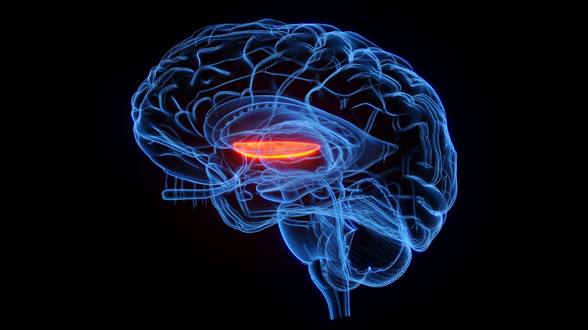
The stimulation was aimed specifically at the right side of a brain structure called the globus pallidus internus (GPI). In a previous study, the authors found that greater connectivity between this region and another structure — the dorsal anterior cingulate gyrus — was linked to more-severe psychotic symptoms in schizophrenia. So, they made the GPI their target.
To date, "there haven't been many successful therapeutic attempts at targeting some of these deeper structures," said Dr. Deepak Sarpal, an associate professor of psychiatry at the University of Pittsburgh School of Medicine who was not involved with the study. Ultrasound waves reach brain regions under the cortex in a less-invasive manner than deep brain stimulation, in which a surgeon implants an electrode in a patient's brain, Sarpal noted.
The researchers are not the only group using transcranial ultrasound to influence the cortico-striatal-pallido-thalamic circuit. Earlier this year, another group stimulated portions of the striatum, which is located directly under the cortex, in patients with schizophrenia. They evaluated hallucinations rather than delusions, finding that the treatment reduced symptoms for at least a week after the procedure.
In this line of research, the targeted nature of transcranial ultrasound presents advantages as a research tool as well as a treatment approach.
"If we find that a specific circuit or a node in a circuit seems to be closely linked to symptoms we're trying to treat, then it will be a matter of understanding what pattern of stimulation may allow us to correct that," Goff said. Qi, Goff and colleagues now plan to continue testing their technique on more patients and to experiment with different doses of ultrasound.
"A lot more work needs to be done," Goff said, "but we're really quite optimistic about its promise."
This article is for informational purposes only and is not meant to offer medical advice.
See how much you know about the most complex organ in the human body with our brain quiz!
Recent research challenges a long-standing idea about how the brain develops: Rather than arising from a single source, the brain may emerge from two distinct populations of cells, the study finds.
"The brain appears as one organ," said study co-author Kyle Loh, an associate professor of developmental biology at Stanford University. "However, we discovered that the front part of the brain and the back part of the brain come from different sources, which are pushed together to create the appearance of a single organ," Loh told Live Science in an email.
The findings, published in September in the journal Nature Neuroscience, not only challenge established models of how the brain forms but may also explain why researchers have struggled to grow certain types of brain cells in the laboratory. The scientists say the work could offer new ways to investigate diseases that affect the brainstem, the "stalk" that connects the brain to the spinal cord.
The human brain is divided into three main regions — called the forebrain, midbrain and hindbrain — that perform different but interconnected functions.
The forebrain, which includes the cerebral hemispheres, is involved in higher-level cognitive processes, such as language, planning and abstract thinking. The midbrain, the uppermost part of the brainstem, connects the forebrain and the hindbrain and acts as a key relay center for sensory information and movement control. The hindbrain — which includes the bottom of the brainstem, along with other structures — also helps to coordinate movement, as well as regulate essential functions such as breathing, sleep and heart rate.
Historically, the hindbrain has been difficult to study. One reason is that scientists have faced persistent challenges in generating functional hindbrain neurons in laboratory conditions.
To help understand why, Loh and his team looked at the early stages of embryonic development in lab mice. They focused on gastrulation, the point when cells begin to organize into structures that will later form different organs. The researchers identified two distinct populations of immature cells that would go on to give rise to nervous system tissue, including the brain.
One cell population expressed a gene called Otx2, which helps to direct the early development of the forebrain and midbrain. The second group of cells expressed the gene Gbx2, which is involved in the development of the midbrain and hindbrain.
The functions of these genes were already known, but their distinct expression in these two groups of cells was revealed by the new research. The cell populations remained distinct as they developed into full-fledged nervous tissue, rather than overlapping.
The team also examined chromatin, the molecular structure that packages DNA tightly inside the nucleus and helps regulate which genes a cell can use. The two cell populations show different chromatin configurations, the team found, which further supports the idea that they follow separate developmental pathways.
That may help to explain why scientists' attempts to grow hindbrain neurons have been largely unsuccessful.
"Previous attempts to make hindbrain neurons likely tried to coax forebrain and midbrain progenitors into hindbrain cells, which our study shows is not possible," study co-author Rayyan Jokhai, a graduate student in Loh's lab, said in a statement.
Using the information uncovered in their experiments, the researchers successfully guided human stem cells to transform into functional hindbrain motor neurons, which are involved in controlling movement. Specifically, these cells produced proteins characteristic of the hard-to-grow hindbrain regions that help control muscles of the face and those involved in swallowing.
In several neurological diseases, including spinal muscular atrophy and amyotrophic lateral sclerosis, motor neurons deteriorate and affected patients can lose their ability to swallow, speak and breathe independently.
"We cannot sample living motor neurons from a patient's hindbrain to watch how these diseases attack these neurons," Loh said. "Growing these hindbrain motor neurons in a dish gives us a way to investigate these diseases." Looking forward, the team aims to use their findings to study these diseases and pinpoint new treatments.
The new findings could also reveal something fundamental about the evolution of vertebrate animals.
The study authors investigated whether the two developmental pathways were unique to humans or reflected a much older evolutionary arrangement. They turned to previously published data on other species — including macaques (Macaca), zebrafish (Danio rerio) and chickens (Gallus gallus domesticus) — and again found two distinct cell populations expressing the two key genes.
And even in acorn worms — a marine invertebrate whose evolution diverged from vertebrates about 550 million years ago — they again found these two distinct groups of cells.
"The Stanford discovery establishes that the deep ancestry of the vertebrate brain is fundamentally modular," said Anna Di Cosmo, a professor of biology at the University of Naples Federico II in Italy who wasn't involved with the study. "The developmental separation between the anterior lineage (forebrain/midbrain) and the posterior lineage (hindbrain) is traceably ancient," Cosmo told Live Science in an email.
Loh's team speculates that these developmental trajectories seen in the womb may suggest that the two cell populations once belonged to two separate nervous systems. "Later in evolution, these two nervous systems may have been pushed together to give rise to the appearance of one integrated brain," Loh said.
However, the study doesn't reveal exactly when the two pathways arose or establish how they integrated. "It is important to emphasize that this is only an idea, and it might change as scientists learn more," Loh said.
See how much you know about the most complex organ in the human body with our brain quiz!
For people with chronic back pain, everyday movements like bending down to tie their shoes or picking up a grocery bag can trigger painful flare-ups. Now, scientists have made a surprising discovery: These episodes may activate a brain network that's associated with our sense of self.
In a new study, the researchers found that movement-induced chronic back pain was tied to increased activity in the brain's default mode network, a group of connected brain regions that lights up during self-reflection and mind wandering. This finding was unexpected because the network typically becomes less active during acute pain, such as transient pain triggered from touching a hot stove or banging your knee on a table.
The new study, published Oct. 5 in the British Journal of Anaesthesia, suggests that chronic-pain flare-ups may involve brain processes beyond those responsible for detecting painful sensations.
Chronic lower back pain, defined as lasting at least three months, is a leading cause of disability worldwide. It's thought that when chronic pain first begins, a network of regions in the brain called the "pain neuromatrix" becomes sensitized so that the pain persists even after the painful stimulus is gone. Although scientists know that chronic pain is associated with changes in brain activity, they don't fully understand what happens when movements trigger sudden increases in that ongoing pain.
To investigate, researchers recruited 114 adults with chronic lower back pain and used functional magnetic resonance imaging (fMRI) to examine their brain activity. fMRI measures changes in blood flow, which acts as an indirect measure of brain activity.
After an initial scan, a researcher helped lift each participant's leg until their back pain increased, held it in position for 10 seconds, and then lowered it. The researchers scanned the participants' brains immediately after this pain-inducing leg movement and compared the results with scans collected beforehand. On average, the test increased the participants' reported pain intensity by 23%.
As expected, during the leg movement, the researchers observed increased blood flow in brain regions involved in processing pain and movement. But they also detected increased activity in key regions of the default mode network, which typically becomes more active when a person is thinking about themselves or their autobiographical memories or just letting their mind wander.
We expected it to be deactivated to the point that we had to triple-check our work to make sure that the results were legit.
Fadel Zeidan, neuroscientist at UC San Diego
Acute pain lowers activity in the default mode network, studies show, presumably because it induces an awareness of something separate from yourself.
"When we saw just the first round of analysis, we're like, 'Did we do it right?'" study co-author Morgan Gianola, a postdoctoral scholar at the University of California, San Diego, told Live Science.
"We did not expect [the default mode network] to be involved at all," said study co-author Fadel Zeidan, a neuroscientist at UC San Diego, told Live Science. "We expected it to be deactivated to the point that we had to triple-check our work to make sure that the results were legit."
The researchers also exposed participants' calves to painful heat — a common method of studying brain activity during acute pain. Unlike the movement-induced back pain, the heat was associated with reduced activity in part of the default mode network, suggesting a different mechanism was at play.
Participants who reported more severe chronic back pain had stronger connectivity between the default mode network and brain regions that process sensations from the back. This suggests that chronic pain may become intertwined with how people perceive themselves and their bodies, the study authors concluded.
"This pain flare has almost become a part of who they are from an egocentric perspective," Zeidan said.
Katherine Martucci, a neuroscientist at Duke University who wasn't involved in the study, praised the experimental design, noting that researchers typically avoid movement during brain scans because it can interfere with the clarity of the scan. This study "nicely gets around" that problem by taking scans before and after the painful motion, Martucci told Live Science in an email.
However, she noted that the observed brain activity could also reflect lingering pain or anxiety following the painful leg raise. In addition, the study couldn't determine whether the altered brain activity observed contributes to chronic pain or develops in response to it.
Nonetheless, Zeidan and colleagues think the findings could eventually inform future treatments for chronic pain. The therapies might address how people relate to their pain, rather than solely targeting the painful sensations themselves. Zeidan, who coincidentally developed chronic back pain while conducting the study, believes that people's past experiences and memories with chronic pain have influenced their brain processes to a point where the pain itself becomes part of who they are.
Zeidan's team is now investigating whether existing approaches, such as mindfulness meditation or cognitive behavioral therapy, can influence the default mode network and help relieve chronic pain. (Cognitive behavior therapy for chronic pain, or CBT-CP, is already a well-established treatment option.)
Martucci cautioned that it's too early to draw conclusions about how these results might be applied in clinical practice. But she said the current findings provide important evidence of how the brain responds to chronic pain flare-ups.
"They do provide solid data on how people with chronic low back pain show important changes in their brain responses after a condition provoking their pain," she said. "The brain imaging data validate their [the participants'] experience of pain in an important and very visible way."
This article is for informational purposes only and is not meant to offer medical advice.
For about a decade, the mainstream understanding has been that psychedelics cause chaos in the brain. Brain networks—the stable patterns of communication that handle vision, attention, or our sense of self, and more—loosen and start talking to each other all at once. EEG traces get noisier and more complex. “Think of the networks of the brain as highways,” says Devon Stoliker, a neuroscientist at Monash University. “Under psychedelics, these highways break down into many different directions.”
In a recent Nature study, Stoliker’s team designed an experiment to test whether these directions are truly chaotic and random. Researchers gathered 62 people who had never taken a psychedelic before, gave them 19 milligrams of psilocybin, a common psychedelic found in certain fungi, and scanned their brains. Then, they used AI to analyze the drugged and sober brain scans. What emerged was not exactly chaos.
Stoliker thinks chaos is not a satisfactory explanation for psychedelics’ effect on the brain. “It never really explained why an individual would have a meaningful experience, why they might have insight, why they might experience clarity, and why these might translate into positive psychological changes,” Stoliker says. To investigate if there are other explanations, he and his colleagues designed a study called PsiConnect.
© Tashi-Delek
Scientists have developed remarkably long, precisely patterned molecular threads that could open new possibilities for designing advanced materials. The scientists also discovered these threads — called supramolecular polymers — can dramatically enhance brain cell activity. 1
Built from self-assembling molecules, the longest threads reach dimensions greater than the diameter of a cell. By molecular standards, they are giant — roughly 100 times more massive than even very large conventional polymers and the largest known proteins in biology. Yet despite their enormous size, scientists can precisely control their lengths and organize them into chemically distinct segments with defined dimensions carrying opposite electrical charges.
That precision also produced an unexpected biological effect. In cell culture studies, the structures enhanced the growth and organization of neurons and increased synapse formation.
The findings introduce a new way to use self-assembly to build highly controlled supramolecular materials with precise physical arrangement of chemical structures. Ultimately, the work could inform materials’ design for a range of applications, including biomaterials for regenerative medicine.
“These structures represent a breakthrough in materials design, and we already discovered one useful application: their superbioactivity toward neurons, which will bring new opportunities in regenerative medicine,” said Northwestern’s Samuel I. Stupp, who led the study. “Typically, when we design a regenerative material, we add biological signals, designed to activate cell receptors. Here, the material itself becomes highly bioactive simply through the precise organization of electrical charges and its dynamic behavior. That introduces a very different way of thinking about how materials can communicate with cells.”
The new study builds on Stupp’s long history working with supramolecular materials, dynamic structures that form when molecules spontaneously organize through weak, reversible interactions. While conventional polymers, like plastics, hold together using strong, permanent chemical bonds, supramolecular polymers are dynamic, allowing their components to move and reconfigure, which generates life-like properties in synthetic systems. Supramolecular polymers exist in all living organisms, where they perform essential functions and hold promise for future soft matter technologies.
That flexible, dynamic nature can be useful for interacting with living cells in biomedical applications, but it also makes the structures difficult to control. Scientists can design the individual molecular building blocks but then struggle to control how long the resulting structures grow and how they organize their chemical components.
Stupp’s team overcame that limitation by using novel designs of molecules and carefully controlling their assembly process. With this strategy, the researchers could dictate the threads’ lengths and keep them uniform. Once the growth stopped, the structures maintained their precise lengths rather than fusing together or rearranging.
“We let these filaments sit in a test tube for months, and they surprisingly didn’t change,” Stupp said. “They remained the same length and retained their segmented structures.”
Stupp and his team traced that unusual ability to a previously unknown process his laboratory calls “self-capping supramolecular polymerization.” As a thread grows, flexible portions at the ends remain highly dynamic and, at proper temperatures, allow new molecules to join in a synchronized manner. Once the available building blocks are consumed, those flexible portions fold over and protect the ends. This effectively shuts down further growth by preventing threads from fusing together or undergoing “Ostwald Ripening,” a process in which smaller structures disappear while larger ones grow.
“To make them longer and longer, we just kept adding new molecules,” Stupp said. “The new molecules stick with strong noncovalent bonds to the growing thread and elongate the assembly, which led to a breakthrough in terms of size control.
“The collective molar mass of their noncovalently bonded monomers reaches billions of daltons, and thus they are ‘giga-assemblies.’ These are the largest supramolecular polymers ever made with this degree of precision. Once the monomer is fully consumed, they finish growing and their dimensions remain stable.”
Approximately the size of one proton or neutron, a dalton is a tiny unit that scientists use to measure the mass of molecules and atoms. More than a billion daltons is enormous by molecular standards. For comparison, the largest common polymers of plastics have molar masses in the range of 10 million daltons, and the largest proteins in nature reach approximately 3 to 5 million daltons.
Researchers can later restart growth by supplying more building blocks. By adding different types of molecules in sequence, the researchers can create precisely defined segments with different chemical properties. In the study, Stupp and his team created threads with positively and negatively charged segments. Because opposites attract, the researchers expected the threads to clump together. Instead, they remained stable in water because the “capping” mechanism prevented the assemblies from fusing together.
To explore how the pattern of electrical charges might interact with living cells, Stupp and his team turned to neurons, which carry a strong negative charge. The team exposed several threads with different designs to cellular cultures containing cortical neurons.
“The first thing we did was make a structure with only positively charged segments, and it basically killed the neurons,” Stupp said. “Then, we tried a structure with only negatively charged segments, and nothing happened because the neurons repelled the materials.”
When Stupp’s team used supramolecular assemblies with randomly mixed positive and negative charges, the neurons didn’t respond in a notable way. But then the team designed a thread with the breakthrough polymerization strategy comprising a small positively charged segment sandwiched between two negatively charged segments of different lengths. Something interesting and unexpected occurred.
The small positive region anchored the thread to the negatively charged surface of a neuron. Repelled by the neuron’s negative charge, the thread’s negative segments remained highly mobile and in constant motion since they could not escape.
“When we used only positively charged segments, they suffocated the neuron and killed it,” Stupp said. “But with only one positively charged segment surrounded by negative ones, it couldn’t do that anymore. And the negative segments, which ordinarily couldn’t get near the neuron, were forced to stay near it because they were anchored.”
Neurons treated with the precisely segmented threads grew longer neurites — the long projections neurons use to communicate with other cells. The treated neurons also formed more elaborate branches, gaining the ability to connect with neighboring cells.
“The neurons matured to an amazing level,” Stupp said. “They grew long and highly branched neurites, which is how they can start making synaptic connections in the brain and in the spinal cord.”
After just seven days, treated neurons were much more active than untreated neurons. When stimulated, treated neurons produced a calcium response — a sign of neuronal activity. Their overall calcium activity was four times greater. And, after 14 days, treated neurons formed more synapses than untreated neurons.
Stupp and his team saw another striking effect in cultures of human neural progenitor cells, which are immature cells that can develop into neurons. Although these cultures typically grow in flat layers, the cells instead organized into raised, three-dimensional clusters connected by axons. This organization resembled aspects of the brain’s white and gray matter, where somas are separated from axons.
Perhaps most surprisingly, the molecular threads produced these effects without carrying biological signals specifically designed to encourage neurons to grow. Stupp posits the negatively charged segments recruit specific proteins already present around the cells that help neurons survive, grow and communicate. These proteins, called neurotrophic factors, have positively charged domains. Stupp’s team also found laminin — an important protein that holds tissues together and has positively charged domains — accumulating along the supramolecular threads near neurons.
Stupp hypothesizes that the interplay between attraction and repulsion causes the threads’ negative segments to rapidly move toward and away from the neurons in a “tapping” motion. Proteins recruited by the negative segments may be drawn toward receptors on the neurons, while electrostatic forces push the segments away. This repeated tapping, occurring on millisecond timescales, could help enhance biological signaling. A live cell movie included in the Science paper has captured this phenomenon.
Beyond neurons, Stupp says the new polymerization strategy could eventually enable scientists to build long molecular structures containing many carefully positioned chemical segments. That could open opportunities to create new materials and devices with features that extend over very long distances.
“We discovered something we had never seen before in chemistry or in biology,” Stupp said. “This work gives us a new level of control over supramolecular materials. In the future, we can imagine creating many segments with different properties. The unknown functions of these structures are likely to be as surprising as the superbioactivity observed on neurons. We’re just beginning to imagine the possibilities.”
The post Supramolecular polymers can dramatically enhance brain cell activity appeared first on Mapping Ignorance.