 Login

Ultrasound waves that jiggle deep brain structures may relieve delusions in schizophrenia, small trial hints
Stimulating the brain with ultrasound could help counter the delusions people experience in schizophrenia, a small trial finds.
The trial involved transcranial ultrasound stimulation, in which a device placed on a patient's head delivers ultrasound waves through the skull and into the brain. Its goal is to change the activity in a targeted brain region.
Delusions are false beliefs that are very difficult to shake; for example, a person might believe they are being stalked when they aren't or think that a major disaster is about to occur without any evidence to suggest so. In the trial, researchers found that a 10-minute session of the ultrasound treatment reduced the severity of delusions for at least a week in patients with schizophrenia or a related disorder.
This targeted ultrasound treatment delivered directly to the brain could avoid some of the full-body effects of antipsychotic medications, such as weight gain and high blood pressure. That said, the new results, published Tuesday (Oct. 20) in the journal npj Acoustics, are a first step and will need further verification in larger trials.
"It's too early to know for certain what the therapeutic benefits will be, but it's very exciting," study co-author Dr. Donald Goff, a professor of psychiatry at the New York University Grossman School of Medicine, told Live Science.
Targeting deep brain structures
The pilot study included 10 patients with either schizophrenia or schizoaffective disorder. In both conditions, patients experience psychotic symptoms, including delusions and hallucinations. Schizoaffective disorder also includes mood symptoms, such as mania.
The researchers interviewed each participant to characterize the content of their delusions, and also documented their cognitive state and experiences with auditory hallucinations — perceptions of sounds that aren't actually occurring. The participants then received both an ultrasound treatment and a placebo treatment, spaced one week apart.
Other noninvasive therapies — such as transcranial magnetic stimulation, which uses magnetic fields — can also stimulate the brain. But the ultrasound technique differs from these therapies because ultrasound waves can penetrate deeper into the brain with greater specificity, study co-author Shy Shoham, a neural engineer at NYU Grossman, told Live Science. He compared the way a transducer directs ultrasound waves toward a given brain region with the way a lens focuses light.
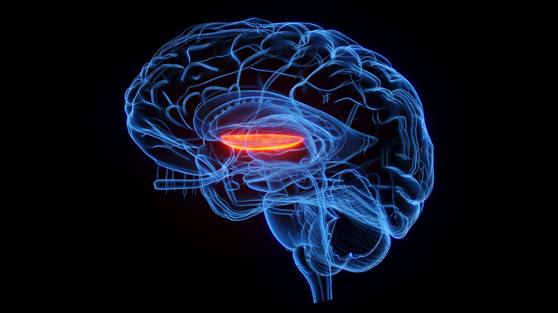
In this case, the researchers focused this lens on a brain network called the cortico-striatal-pallido-thalamic circuit, which includes structures underneath the brain's outer cortex. The relationship between this circuit and schizophrenia symptoms is still somewhat unclear, partly because its deeper components are hard to study in adequate resolution with MRI, said co-author Dr. Wei Qi, a clinical assistant professor of psychiatry at NYU Grossman. However, some past studies have suggested that this network plays a part in schizophrenia.
Much of what scientists know about the network comes from research in movement disorders, such as Parkinson's disease, in which there is an imbalance between excitation and inhibition in the neurons that control movement. A similar dynamic could help to explain symptoms of schizophrenia, the scientists think.
"Our hypothesis is that, in schizophrenia, there is an imbalance between excitation and inhibition regarding what in the environment is actually important," Qi told Live Science. "The failure to inhibit irrelevant signals could be the basis of psychosis."
"Quite optimistic"
The researchers surveyed the study participants after each treatment, asking about the degree of belief each person had in their delusions and the amount of stress they caused. Compared with the placebo treatment, the ultrasound stimulation was associated with less-severe delusions over the following week. There was no significant change in patients' auditory hallucinations or overall cognition, though.
The ultrasound therapy came with mild side effects, such as fatigue and headache. Some patients had transient increases in delusions, which Qi attributed to normal fluctuations in symptoms that are expected in schizophrenia. These outcomes were just as common after the placebo treatment, so they may not be related to the ultrasound specifically.
The stimulation was aimed specifically at the right side of a brain structure called the globus pallidus internus (GPI). In a previous study, the authors found that greater connectivity between this region and another structure — the dorsal anterior cingulate gyrus — was linked to more-severe psychotic symptoms in schizophrenia. So, they made the GPI their target.
To date, "there haven't been many successful therapeutic attempts at targeting some of these deeper structures," said Dr. Deepak Sarpal, an associate professor of psychiatry at the University of Pittsburgh School of Medicine who was not involved with the study. Ultrasound waves reach brain regions under the cortex in a less-invasive manner than deep brain stimulation, in which a surgeon implants an electrode in a patient's brain, Sarpal noted.
The researchers are not the only group using transcranial ultrasound to influence the cortico-striatal-pallido-thalamic circuit. Earlier this year, another group stimulated portions of the striatum, which is located directly under the cortex, in patients with schizophrenia. They evaluated hallucinations rather than delusions, finding that the treatment reduced symptoms for at least a week after the procedure.
In this line of research, the targeted nature of transcranial ultrasound presents advantages as a research tool as well as a treatment approach.
"If we find that a specific circuit or a node in a circuit seems to be closely linked to symptoms we're trying to treat, then it will be a matter of understanding what pattern of stimulation may allow us to correct that," Goff said. Qi, Goff and colleagues now plan to continue testing their technique on more patients and to experiment with different doses of ultrasound.
"A lot more work needs to be done," Goff said, "but we're really quite optimistic about its promise."
This article is for informational purposes only and is not meant to offer medical advice.
See how much you know about the most complex organ in the human body with our brain quiz!